The discovery of the cell, the fundamental unit of life, stands as a monumental achievement in the field of biology. Through centuries of relentless inquiry and scientific curiosity, numerous visionaries and scientists have contributed to unraveling the intricate world of cells. This article explores the captivating history of cell discovery, highlighting the key individuals, important concepts, and groundbreaking experiments that paved the way for our modern understanding of cellular biology.

In the late 17th century, the Dutch tradesman and scientist Antonie van Leeuwenhoek emerged as a pioneer in the field of microscopy. Armed with simple yet ingenious lens grinding techniques, van Leeuwenhoek constructed powerful microscopes capable of observing the microscopic realm. In 1674, he became the first person to witness living cells under a microscope, peering at a variety of specimens including bacteria, sperm cells, and red blood cells. His meticulous observations, meticulously recorded in his letters to the Royal Society of London, shed light on the existence of a previously unseen world of minute organisms.
Robert Hooke and the Beginnings of Cell Theory:
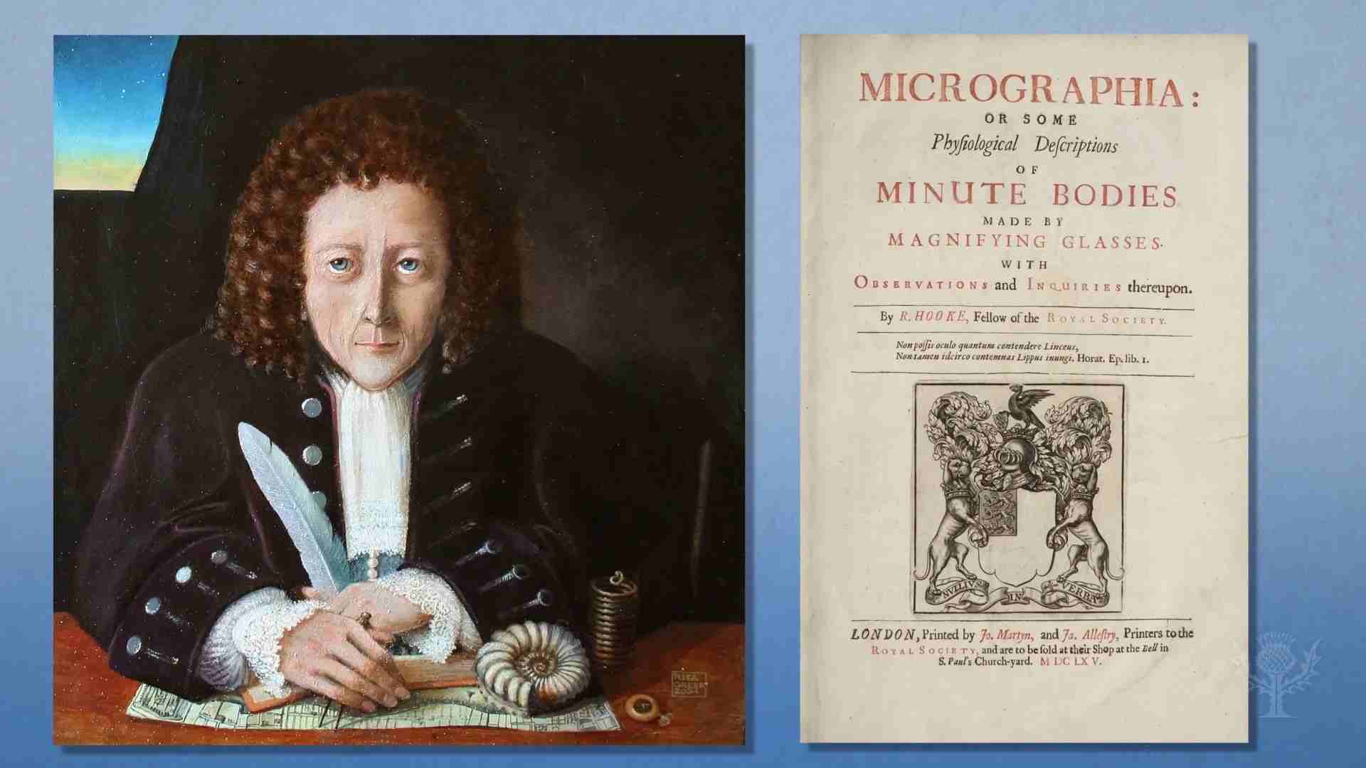
In 1665, Robert Hooke, an English natural philosopher, published his groundbreaking work, “Micrographia.” Within its pages, Hooke described his observations of thin slices of cork, which he termed “cells” due to their resemblance to small compartments or prison cells. Hooke’s discovery laid the foundation for the development of cell theory, the concept that all living organisms are composed of cells.
In the 19th century, two scientists, Matthias Jakob Schleiden and Theodor Schwann, played pivotal roles in formulating the modern cell theory. Schleiden, a German botanist, proposed that plants were composed of cells, while Schwann, a German physiologist, extended this concept to animals. Together, their work demonstrated that cells were the fundamental units of all living organisms, and their research laid the foundation for the development of modern cell biology.
Another key figure in the history of cell discovery is Rudolf Virchow, a German pathologist. In 1855, Virchow proposed the concept of cellular reproduction through his influential phrase, “omnis cellula e cellula” or “every cell originates from another existing cell.” This principle emphasized that cells arise from pre-existing cells, challenging the previously held notion of spontaneous generation.
Over the years, advancements in microscopy, biochemistry, and molecular biology have further enhanced our understanding of cells. Scientists such as Robert Brown, who discovered the cell nucleus in 1831, and James Watson and Francis Crick, who elucidated the structure of DNA in 1953, have contributed significantly to our comprehension of cell function and genetics.
Today, the study of cells continues to evolve, with ongoing research in areas like stem cell biology, cellular signaling, and genetic engineering. Prominent research institutions, including the Massachusetts Institute of Technology (MIT), the Max Planck Institute, and the National Institutes of Health (NIH), are at the forefront of groundbreaking research on cells.
Cell Theory:

Cell theory, one of the cornerstones of modern biology, revolutionized our understanding of life and its fundamental units. Developed through the contributions of several prominent scientists, the theory lays the groundwork for comprehending the complex structures and functions of organisms. This extensive article explores the fascinating history of cell theory, highlighting the key individuals, pivotal experiments, and groundbreaking concepts that have shaped our knowledge of cells.
In the early 19th century, the German botanist Matthias Jakob Schleiden made significant strides toward formulating cell theory. Through his meticulous investigations of plant tissues, Schleiden proposed that plants were composed of discrete units called “cells”. His observations, documented in his influential work “Contributions to Phytogenesis,” laid the foundation for the concept that cells were the building blocks of plant structures.
Building upon Schleiden’s work, the German physiologist Theodor Schwann expanded the scope of cell theory to encompass animal tissues. In his seminal publication “Microscopic Investigations on the Accordance in Structure and Growth of Animals and Plants,” Schwann established that animal tissues also consisted of individual cells. He postulated that cells were not only the structural units of living organisms but also the sites of vital physiological processes.
A pivotal figure in the advancement of cell theory, the German physician and pathologist Rudolf Virchow introduced the concept of cellular reproduction. Through his groundbreaking research, Virchow proposed the famous phrase, “omnis cellula e cellula” or “every cell arises from a pre-existing cell.” This fundamental principle underscored the idea that cells divide and give rise to new cells, challenging the notion of spontaneous generation and emphasizing the continuity of life.
Key Tenets of Cell Theory:
The development of cell theory led to the establishment of its fundamental principles, which remain integral to our understanding of cells and living organisms. These key tenets can be summarized as follows:
- All living organisms are composed of cells: Cell theory asserts that cells are the basic structural and functional units of all living organisms, from single-celled bacteria to complex multicellular organisms.
- Cells are the building blocks of life: Cells are responsible for the organization, growth, and development of organisms. They carry out essential functions such as metabolism, reproduction, and response to stimuli.
- Cells arise from pre-existing cells: The principle of cellular reproduction states that cells only arise from pre-existing cells through the process of cell division. This concept, first proposed by Virchow, forms the basis of modern cell biology.
Since its inception, cell theory has undergone significant advancements, facilitated by technological innovations and research breakthroughs. Modern tools such as advanced microscopy techniques, genetic sequencing, and molecular biology methods have unraveled the intricate details of cellular structure and function.
Prominent research institutions and organizations, including the Max Planck Institute for Molecular Cell Biology and Genetics in Germany, the Harvard Medical School in the United States, and the European Molecular Biology Laboratory (EMBL), have played vital roles in pushing the boundaries of cell theory. Their efforts have contributed to our understanding of cellular processes, intracellular signaling, organelle function, and the regulation of gene expression.
Robert Hooke and His Monumental Contributions to the Study of Cells

Robert Hooke, an influential English scientist and polymath of the 17th century, played a pivotal role in the development of cell biology. His groundbreaking observations and investigations laid the foundation for our understanding of cells and their significance in the world of biology. This extensive article delves into the life and work of Robert Hooke, highlighting his key discoveries, notable achievements, and the lasting impact of his contributions to the study of cells.
Born in 1635 in Freshwater, Isle of Wight, England, Hooke exhibited a keen interest in scientific pursuits from an early age. He began his studies at Westminster School and later joined Christ Church, Oxford, where he worked as an assistant to the renowned physicist Robert Boyle. Hooke’s exceptional skills in mechanics, microscopy, and experimental design set the stage for his groundbreaking contributions to the field of biology.
In 1665, Hooke published his monumental work, “Micrographia”, which catapulted him to scientific fame. This masterpiece of scientific literature showcased his meticulous observations made using a compound microscope of his own design. The book not only provided detailed illustrations of various objects viewed through the microscope but also introduced the term “cell” to describe the tiny structures he observed in cork and other plant tissues.
Hooke’s Observations and the Discovery of Cells:
In “Micrographia,” Hooke examined a thin slice of cork under his microscope and was captivated by the intricate network of small, box-like structures he observed. These structures resembled the small compartments or prison cells of a monastery, leading Hooke to coin the term “cells”. His revolutionary discovery challenged the prevailing belief that living organisms were made up of a homogeneous substance and instead proposed that they consisted of discrete, structural units.
Hooke’s curiosity extended far beyond cork. In “Micrographia,” he documented his observations of various specimens, including fleas, mites, fungi, and plant tissues. His meticulous illustrations and descriptions provided a comprehensive account of the microscopic world, captivating readers and inspiring generations of scientists to delve deeper into the realms of cellular biology.
Hooke’s work on cells marked a crucial turning point in the field of biology. His observations and descriptions laid the foundation for the development of cell theory, which posits that cells are the fundamental units of life. Hooke’s term “cell” became an integral part of scientific vocabulary and is still widely used today.
Hooke’s other contributions were equally significant. He made pioneering advancements in the fields of physics, astronomy, and engineering. His investigations of elasticity led to Hooke’s Law, which describes the relationship between the force applied to an elastic object and the resulting deformation.
Robert Hooke’s work had a profound influence on his contemporaries and future generations of scientists. His revolutionary ideas and keen observational skills inspired scientists such as Antonie van Leeuwenhoek, who further advanced the field of microscopy, and Matthias Jakob Schleiden and Theodor Schwann, who developed cell theory based on Hooke’s initial observations.
Today, Hooke’s legacy is honored through various scientific societies, such as the Royal Society of London, of which he was a founding member and served as curator of experiments. His contributions continue to inspire researchers worldwide, and his spirit of curiosity and innovation serves as a guiding light for the scientific community.
Matthias Jakob Schleiden: Pioneering the Understanding of Cells and Plant Structure

Matthias Jakob Schleiden, a renowned German botanist of the 19th century, made significant contributions to the study of cells and plant biology. His pioneering research and observations laid the groundwork for our understanding of the cellular nature of plants. This comprehensive article delves into the life and work of Matthias Jakob Schleiden, highlighting his key discoveries, notable achievements, and the enduring impact of his contributions to the field of biology.
Born on April 5, 1804, in Hamburg, Germany, Matthias Jakob Schleiden displayed an early interest in natural sciences. He pursued his education at the University of Jena and later University of Berlin, where he studied medicine and botany. These formative years marked the beginning of Schleiden’s illustrious career and his lifelong passion for understanding plant structure and organization at the cellular level.
In 1838, Schleiden published his influential work, “Contributions to Phytogenesis”, which revolutionized the study of plants and cells. In this seminal publication, he put forth the revolutionary idea that plant tissues were composed of individual units known as “cells”. Schleiden’s concept challenged the prevailing belief that plants were homogeneous structures and proposed that cells were the fundamental building blocks of plant tissues.
Schleiden’s research focused primarily on plant cells and their role in the development and function of organisms. He investigated various plant tissues and meticulously documented his observations using microscopes. His meticulous illustrations and descriptions provided valuable insights into the diverse structures and functions of plant cells, further emphasizing their significance in the study of botany.
Contributions to Cell Theory:
Schleiden’s work laid the foundation for the formulation of cell theory, which proposed that cells were the basic units of all living organisms. His observations of plant cells inspired subsequent scientists, including Theodor Schwann in the study of animal cells. Together, Schleiden and Schwann established the concept that cells were the building blocks of all living organisms, thereby revolutionizing the field of biology.
Schleiden maintained a close collaboration with Theodor Schwann, a German physiologist, in the formulation of cell theory. They shared ideas and findings, exchanging scientific correspondence to advance their research. This fruitful collaboration resulted in the integration of their work into a unified theory, solidifying the concept that cells were the fundamental units of both plants and animals.
Matthias Jakob Schleiden’s work significantly advanced our understanding of plant structure and the importance of cells in the biological world. His contributions provided a crucial foundation for the development of modern botany and cell biology. Schleiden’s insights and concepts, such as the significance of cell specialization in plant tissues, continue to shape our understanding of plant biology to this day.
Schleiden’s groundbreaking work earned him recognition and accolades within the scientific community. In 1845, he was appointed as a professor of botany at the University of Dorpat in present-day Estonia. Later, he served as a professor at the University of Jena and the University of Bonn, where he continued to contribute to botanical research and mentor future generations of scientists.
Robert Brown and the Revolution of Cell Nucleus:

Robert Brown, a distinguished Scottish botanist of the 19th century, made groundbreaking discoveries that transformed our understanding of cells and their structure. His meticulous observations and insightful analyses brought to light the existence of a central structure within cells, now known as the cell nucleus. This comprehensive article explores the life and work of Robert Brown, highlighting his key contributions, significant findings, and the lasting impact of his discoveries in the field of cellular biology.
Robert Brown was born on December 21, 1773, in Montrose, Scotland. He developed an early interest in natural history and pursued his passion for botany. Brown’s passion for scientific exploration led him to embark on several expeditions, including one aboard the HMS Investigator during Captain Matthew Flinders’ voyage to Australia.
In the early 19th century, while examining plant cells under a microscope, Robert Brown made a profound discovery. In 1831, he observed a peculiar structure at the center of plant cells, which he termed the “nucleus”. Brown documented his findings in a paper titled “Observations on the Organs and Mode of Fecundation in Orchideae and Asclepiadeae,” marking a significant milestone in the understanding of cellular biology.
The Significance of the Cell Nucleus:
Brown’s discovery of the cell nucleus revolutionized the field of cellular biology. The nucleus, located within the cytoplasm of cells, was revealed to be a distinct and crucial component. Brown’s observations laid the foundation for further research into the functions and characteristics of the nucleus, leading to significant advancements in our understanding of cell division, genetic material, and cellular reproduction.
Apart from his work on cells, Robert Brown made significant contributions to the field of botany, particularly in plant classification and taxonomy. His detailed investigations and careful analyses of plant specimens led to the development of new classification systems and the identification of numerous plant species. Brown’s comprehensive botanical research was instrumental in expanding our knowledge of plant diversity.
Robert Brown’s significant contributions to science were widely recognized during his lifetime. In 1821, he was elected as a fellow of the prestigious Royal Society of London. He served as the President of the Linnean Society of London in 1849 and received numerous accolades for his scientific achievements.
Robert Brown’s discoveries laid the groundwork for further exploration and understanding of cellular biology. His identification of the cell nucleus was a crucial step toward elucidating the intricate mechanisms underlying cell function and reproduction. Brown’s observations inspired subsequent generations of scientists, including Theodor Schwann, Matthias Jakob Schleiden, and Rudolf Virchow, who expanded upon his work and contributed to the formulation of cell theory.
Brown’s discoveries were made possible through advancements in microscopy, which allowed for more detailed observations of cellular structures. The development of powerful microscopes, such as the compound microscope, improved resolution and magnification, enabling scientists to delve deeper into the microscopic world of cells.
Brown’s findings continue to inspire ongoing research and discoveries in the field of cellular biology. Scientists today employ advanced techniques such as electron microscopy, fluorescence imaging, and molecular biology to explore the intricate details of cellular structures and functions. Research institutions and organizations around the world, including the National Institutes of Health (NIH) in the United States and the Max Planck Institute in Germany, remain at the forefront of cell biology research and continue to build upon the foundation laid by Robert Brown. These institutions, along with many others, strive to uncover the mysteries of cell biology, investigating topics such as cell signaling, organelle dynamics, and the interplay between genetics and cellular function.
Brown’s legacy extends beyond his groundbreaking discoveries. His meticulous approach to scientific inquiry, attention to detail, and dedication to expanding human knowledge serve as a source of inspiration for aspiring scientists. His work exemplifies the power of observation and the profound impact that a single individual can have on advancing our understanding of the natural world.
Rudolf Virchow: The Revolutionary Contributions to Cellular Pathology and Cellular Reproduction

Rudolf Virchow, a prominent German physician and pathologist of the 19th century, made groundbreaking discoveries that forever transformed our understanding of cells and their role in human health. Through his pioneering work in cellular pathology and cellular reproduction, Virchow revolutionized the field of medicine and left an indelible mark on the study of cells. This extensive article delves into the life and work of Rudolf Virchow, highlighting his key contributions, notable achievements, and the enduring impact of his discoveries in the field of cellular biology.
Rudolf Ludwig Carl Virchow was born on October 13, 1821, in Schivelbein, Prussia (now Świdwin, Poland). Virchow pursued his medical education at the University of Berlin, where he demonstrated a remarkable aptitude for scientific research and a deep commitment to improving medical practices.
Virchow’s most influential contribution to the field of biology was his establishment of cellular pathology. In his groundbreaking work, “Die Cellularpathologie in ihrer Begründung auf physiologische und pathologische Gewebelehre” (Cellular Pathology as Based upon Physiological and Pathological Histology), published in 1858, Virchow emphasized the cellular origin of diseases. He proposed that diseases arise from abnormalities within cells, challenging the prevailing theories that attributed diseases to imbalances in body fluids or organs.
Virchow’s principle of omnis cellula e cellula (“every cell originates from a pre-existing cell”) further solidified the notion that cellular processes are at the core of both health and disease. His research paved the way for a deeper understanding of cellular mechanisms and laid the foundation for the development of modern pathology.
Cellular Reproduction: Challenging Spontaneous Generation:
Building on the concept of cellular pathology, Virchow made significant contributions to our understanding of cellular reproduction. He refuted the long-standing belief in spontaneous generation, which suggested that living organisms could arise spontaneously from non-living matter. Virchow’s observations and experiments provided evidence that cells only arise from pre-existing cells through the process of cell division.
Virchow’s work on cellular reproduction and his principle of omnis cellula e cellula underscored the continuity of life and challenged earlier notions of spontaneous generation. His discoveries were instrumental in establishing a fundamental principle of modern biology and laid the groundwork for further investigations into cell division and genetic inheritance.
Impact on Medical and Social Reforms:
Beyond his scientific contributions, Virchow’s work had far-reaching implications for medical and social reforms. He championed the idea that social and economic conditions played a significant role in determining the health of populations. Virchow advocated for improved sanitation, access to clean water, and social justice, recognizing that poverty and inequality were underlying factors contributing to disease.
Virchow’s tireless efforts in public health and his commitment to social reform earned him recognition as a prominent figure in both medicine and politics. He served as a member of the Prussian House of Deputies and was a vocal advocate for public health policies that aimed to address the root causes of disease.
Rudolf Virchow’s contributions to the understanding of cells and their role in disease have left an enduring legacy in the fields of medicine and biology. His emphasis on cellular pathology revolutionized medical diagnostics and laid the groundwork for personalized medicine. Virchow’s principles continue to shape the practice of pathology, with his ideas informing modern techniques such as molecular diagnostics and genetic profiling.
Virchow’s commitment to scientific rigor and meticulous observation set a high standard for future researchers. His emphasis on the importance of studying cells at the microscopic level paved the way for advancements in microscopy techniques and the development of staining methods that enhanced the visualization of cellular structures.
Virchow’s work continues to inspire ongoing research and discoveries in the field of cellular biology. His principles and findings have provided a solid foundation for the study of cell biology, cellular pathology, and cellular reproduction. Scientists and researchers at esteemed institutions and organizations worldwide, such as the Max Planck Institute for Molecular Cell Biology and Genetics in Germany and the National Institutes of Health (NIH) in the United States, build upon Virchow’s legacy as they investigate the intricacies of cellular processes and their implications for human health.
Virchow’s influence extends beyond the scientific realm. He played a crucial role in bridging the gap between science and society, emphasizing the importance of addressing social determinants of health and advocating for reforms that aimed to improve public health. His holistic approach to medicine and his recognition of the interconnectedness of biological and social factors continue to inspire researchers, physicians, and policymakers in their efforts to create a healthier and more equitable world.
Theodor Schwann: Pioneering Cell Theory

Theodor Schwann, a prominent German physiologist and histologist of the 19th century, made groundbreaking discoveries that revolutionized our understanding of cells and their vital role in living organisms. Through his meticulous research and innovative experiments, Schwann played a pivotal role in formulating cell theory and expanding our knowledge of animal cells. This extensive article explores the life and work of Theodor Schwann, highlighting his key contributions, significant achievements, and the enduring impact of his discoveries in the field of cellular biology.
Theodor Schwann was born on December 7, 1810, in Neuss, Germany. After completing his medical studies at the University of Bonn and University of Würzburg, Schwann embarked on a remarkable scientific career that would shape the course of cellular biology.
Cell Theory and Animal Cells:
Schwann’s most significant contribution to science was his instrumental role in the formulation of cell theory, which established that cells were the basic units of all living organisms. In 1838, Schwann published his groundbreaking work, “Microscopic Investigations on the Accordance in Structure and Growth of Animals and Plants”, where he detailed his experiments and observations.
Building upon the work of Matthias Jakob Schleiden, who focused on plant cells, Schwann extended cell theory to include animal tissues. He proposed that animal tissues were also composed of individual units called cells, similar to plant tissues. Schwann’s observations challenged the prevailing notion that animals were structurally and fundamentally different from plants, laying the foundation for a unified understanding of cellular biology.
Schwann and Cellular Metabolism:
In addition to his work on cell theory, Schwann made significant contributions to the understanding of cellular metabolism. He conducted experiments investigating the process of digestion, studying the action of gastric juices on food. Schwann’s research led him to propose that digestion was a chemical process occurring within cells, further highlighting the central role of cells in the vital functions of organisms.
Schwann’s research on cell theory paved the way for further investigations into cellular reproduction. In collaboration with Matthias Jakob Schleiden, he explored the process of cell division, aiming to understand how new cells were formed. Their combined efforts laid the groundwork for the principle of cellular reproduction, which states that cells arise only from pre-existing cells.
Theodor Schwann’s contributions to the understanding of cells and cell theory were widely recognized and celebrated. In 1845, he was awarded the Copley Medal by the Royal Society of London for his exceptional achievements in physiology. Schwann’s work laid the foundation for modern cell biology, influencing generations of scientists in their pursuit of unraveling the mysteries of cellular life.
Schwann’s discoveries continue to shape our understanding of cells and their vital functions in living organisms. His work provided a springboard for subsequent advancements in cell biology, including the exploration of organelles, cellular processes, and the intricate mechanisms of cellular communication.
Today, researchers at esteemed institutions and organizations, such as the Max Planck Institute for Medical Research and the National Institutes of Health (NIH), are at the forefront of cell biology research. Their investigations into cellular structure, function, and molecular interactions build upon the foundation laid by Schwann and contribute to our understanding of complex biological processes.
Antonie van Leeuwenhoek: Microscopic World and the Discovery of Cells

Antonie van Leeuwenhoek, a visionary Dutch scientist and tradesman of the 17th century, made remarkable advancements in microscopy and played a crucial role in unraveling the mysteries of the microscopic realm. Through his groundbreaking observations, van Leeuwenhoek became the first person to witness and document living cells, laying the foundation for our understanding of the microscopic world and the fundamental units of life. This extensive article explores the life and work of Antonie van Leeuwenhoek, highlighting his key contributions, notable achievements, and the enduring impact of his discoveries in the field of cellular biology.
Antonie van Leeuwenhoek was born on October 24, 1632, in Delft, Netherlands. Though primarily a tradesman, he possessed an insatiable curiosity and an unwavering dedication to scientific inquiry. Van Leeuwenhoek’s interest in microscopy was ignited when he acquired a single-lens microscope and began to explore the unseen world.
The Discoveries of Microorganisms:
Van Leeuwenhoek’s meticulous observations of various specimens using his self-designed microscopes brought to light an astonishing array of microorganisms. In 1674, he became the first person to document the existence of bacteria by closely examining samples of dental plaque. His observations of these tiny, single-celled organisms challenged prevailing notions of the time and opened up a new field of scientific inquiry.
Observation of Living Cells:
Van Leeuwenhoek’s most groundbreaking achievement came in his observations of living cells. By skillfully grinding and polishing lenses, he constructed microscopes with unprecedented magnification power. In 1676, he turned his lenses toward blood cells, becoming the first person to witness and describe red blood cells and their characteristic discoid shape. He also observed sperm cells, providing valuable insights into reproductive biology.
Van Leeuwenhoek maintained a fruitful correspondence with the prestigious Royal Society of London, sharing his remarkable observations and discoveries. His detailed letters, meticulously written in Dutch, were translated and published in the Society’s journal, “Philosophical Transactions.” These letters revealed the intricate world of cells, bacteria, and microorganisms, captivating the scientific community.
Van Leeuwenhoek’s microscopes were a testament to his ingenuity and craftsmanship. His microscopes featured a tiny glass bead as a lens, which allowed for remarkably high magnification. Van Leeuwenhoek’s ability to meticulously grind and polish lenses enabled him to achieve unprecedented clarity and resolution in his observations, making him a pioneer in the field of microscopy.
Antonie van Leeuwenhoek’s discoveries revolutionized our understanding of the microscopic world and the existence of cells. His meticulous observations and precise documentation laid the foundation for modern cellular biology and paved the way for subsequent advancements in the field. Van Leeuwenhoek’s work inspired generations of scientists, including renowned figures such as Robert Hooke and Matthias Jakob Schleiden, who further expanded our understanding of cells and their structures.
Today, microscopy remains an essential tool in the study of cells and microorganisms. Advanced techniques, such as electron microscopy and fluorescence microscopy, have allowed scientists at prestigious research institutions like the Max Planck Institute and the National Institutes of Health (NIH) to explore the intricacies of cellular structures and functions with unparalleled detail.
Cell Structure:

Key Components of Cell Structure:
- Cell Membrane: The outer boundary of a cell, composed of a lipid bilayer, which regulates the exchange of materials between the cell and its environment.
- Cytoplasm: The gel-like substance filling the cell, where various organelles and cellular structures are suspended.
- Nucleus: The central control center of the cell, containing genetic material in the form of DNA and responsible for regulating cellular activities.
- Organelles: Specialized structures within the cell that perform specific functions, such as mitochondria for energy production, endoplasmic reticulum for protein synthesis, and Golgi apparatus for protein modification and sorting.
- Cytoskeleton: A network of protein filaments that provides structural support and facilitates cell movement and shape maintenance.
- Chromosomes: Structures within the nucleus that contain DNA, the genetic blueprint of an organism, and play a crucial role in cell division and inheritance.
The advancement of imaging techniques has greatly enhanced our ability to explore cell structure in greater detail. Techniques such as light microscopy, electron microscopy, and confocal microscopy have allowed scientists to visualize cells and their components with high resolution, enabling the study of cellular structures and interactions.
Numerous scientists have made significant contributions to the understanding of cell structure. Notable names include Ernst Ruska, who invented the electron microscope, and Christian de Duve, who discovered lysosomes and peroxisomes. Institutions like the Howard Hughes Medical Institute and the European Molecular Biology Laboratory have been at the forefront of cutting-edge research on cell structure.
Ongoing research continues to unravel new aspects of cell structure. Concepts such as cellular compartmentalization, cellular signaling, and nanoscale organization are areas of active investigation. Scientists are exploring the three-dimensional architecture of cells and the interplay between different cellular components to gain a comprehensive understanding of how cells function and respond to stimuli.
Seeing Inside Cells:

The quest to see inside cells began centuries ago with the advent of microscopy. Pioneers such as Robert Hooke and Antonie van Leeuwenhoek utilized early microscopes to observe cells, laying the foundation for the field of cell biology. Hooke’s observations of cork cells and Leeuwenhoek’s investigations of various specimens opened up a new world of microscopic exploration.
Light Microscopy: Revealing Cellular Structures:
The development of light microscopy revolutionized our ability to visualize cellular structures. In the 19th century, Ernst Abbe and Carl Zeiss made significant advancements in lens technology, improving resolution and enabling the visualization of finer details within cells. The introduction of staining techniques by scientists like Golgi and Nissl further enhanced the contrast and clarity of cellular components, facilitating the identification of organelles and subcellular structures.
Electron Microscopy: Unveiling the Nanoscale World:
A major leap forward came with the invention of the electron microscope. In the 1930s, Max Knoll and Ernst Ruska collaborated to develop the first electron microscope, which used a beam of electrons instead of light to visualize cellular structures with unprecedented resolution. This breakthrough technology allowed scientists to peer into the nanoscale world, revealing intricate details of cellular organelles and molecular structures.
Fluorescence microscopy introduced the ability to visualize specific molecules and processes within cells. By using fluorescent dyes and fluorescent proteins such as Green Fluorescent Protein (GFP), researchers can selectively label cellular components and track their movements in real-time. The work of scientists like Roger Y. Tsien and Stefan Hell led to advancements in super-resolution fluorescence microscopy, allowing scientists to surpass the diffraction limit and visualize cellular structures at the molecular level.
The development of techniques for live cell imaging has revolutionized our understanding of cellular dynamics. Through time-lapse imaging and the use of confocal microscopy and total internal reflection fluorescence microscopy (TIRF), researchers can observe cellular processes in real-time. This has provided insights into phenomena such as cell division, cell migration, and intracellular signaling, shedding light on the mechanisms underlying cellular behavior.
Cutting-Edge Technologies and Institutions:
Prominent institutions and organizations around the world are at the forefront of cutting-edge research on cellular imaging. Places like the Janelia Research Campus and the Wellcome Trust Sanger Institute house state-of-the-art imaging facilities and bring together multidisciplinary teams of scientists to explore the intricacies of cellular life.
Advancements in technology continue to push the boundaries of cellular imaging. Concepts such as correlative microscopy, single-molecule imaging, and cryo-electron microscopy are opening new frontiers in understanding cellular processes with unprecedented detail. Additionally, the integration of imaging techniques with computational analysis and machine learning holds tremendous potential for extracting complex information from cellular imaging data.
Key Dates:

1665: Robert Hooke and the Cork Cells:
In 1665, Robert Hooke, an English scientist, observed and documented cells for the first time. Using a compound microscope, he examined a thin slice of cork and observed a network of small, box-like structures. Hooke named these structures “cells”, drawing an analogy to the small compartments or prison cells of a monastery. This marked the first recorded observation of cells and laid the foundation for the study of cellular biology.
1674: Antonie van Leeuwenhoek and the Microscopic World:
In 1674, the Dutch scientist Antonie van Leeuwenhoek made significant contributions to cell discovery. He constructed powerful single-lens microscopes, enabling him to observe a vast array of microorganisms. Van Leeuwenhoek’s meticulous observations revealed the existence of bacteria, protozoa, and other microscopic organisms, expanding our understanding of the diverse microscopic world within cells.
1838: Matthias Jakob Schleiden and Plant Cells:
In 1838, the German botanist Matthias Jakob Schleiden proposed that plants were composed of individual units called “cells”. He investigated various plant tissues and documented his observations, highlighting the structural and functional importance of cells in plant biology. Schleiden’s work, along with his collaboration with Theodor Schwann, formed the basis of cell theory, which revolutionized our understanding of cellular biology.
1839: Theodor Schwann and Animal Cells:
In 1839, the German physiologist Theodor Schwann extended cell theory to animal tissues. He proposed that animal tissues, like plant tissues, were also composed of cells. Schwann’s research and collaboration with Schleiden led to the formulation of cell theory, which postulated that cells are the fundamental units of all living organisms. Schwann’s work played a crucial role in unifying the understanding of cells across different organisms.
1855: Rudolf Virchow and Cellular Reproduction:
In 1855, the German physician Rudolf Virchow introduced the concept of cellular reproduction. He proposed that cells arise only from pre-existing cells, challenging the prevailing theory of spontaneous generation. Virchow’s principle of omnis cellula e cellula (“every cell originates from a pre-existing cell”) further solidified the understanding of cellular reproduction, highlighting the continuity of life and the importance of cell division in growth and development.
1931: Ernst Ruska and the Electron Microscope:
In 1931, German physicist Ernst Ruska developed the first practical electron microscope. This revolutionary invention utilized a beam of electrons instead of light, allowing for higher resolution and magnification than traditional microscopes. The electron microscope enabled scientists to explore cellular structures at the nanoscale level, unveiling the intricate details of organelles and molecular components within cells.
Late 20th Century: Advancements in Imaging Technologies:
The latter half of the 20th century saw remarkable advancements in imaging technologies. Techniques such as fluorescence microscopy, confocal microscopy, and super-resolution microscopy allowed scientists to visualize specific molecules, track dynamic processes within cells, and surpass the limitations of traditional microscopy.
2000s: Advances in Live Cell Imaging and Molecular Probes:
In the 21st century, there have been significant advancements in live cell imaging techniques. Scientists developed innovative methods to capture and study cellular processes in real-time, including time-lapse imaging, total internal reflection fluorescence microscopy (TIRF), and single-molecule imaging. The introduction of fluorescent molecular probes, such as green fluorescent protein (GFP) and its variants, enabled researchers to label specific cellular components and track their movements and interactions within live cells.
Modern Era: Integration of Imaging and Computational Analysis:
In recent years, the integration of imaging techniques with computational analysis and machine learning has opened new avenues for understanding cellular biology. Researchers can extract valuable quantitative information from complex imaging data, analyze cellular dynamics, and unravel intricate relationships within cellular structures and functions.
In conclusion
The quest for knowledge and discovery has led to remarkable advancements in various fields of study. Throughout history, numerous scientists and researchers have dedicated their lives to unraveling the mysteries of the world, making groundbreaking discoveries that have shaped our understanding of the natural world and propelled human progress.
The individuals mentioned in this article, including Robert Hooke, Antonie van Leeuwenhoek, Matthias Jakob Schleiden, Theodor Schwann, Rudolf Virchow, and Ernst Ruska, among others, are just a few of the brilliant minds who have significantly contributed to our knowledge of cells and cellular biology. Their keen observations, meticulous experiments, and innovative inventions have paved the way for the establishment of cell theory, the exploration of cellular structures, and the unveiling of the intricate processes that occur within cells.
These pioneers and their groundbreaking work have shaped the foundation of modern biology and continue to inspire generations of scientists to push the boundaries of knowledge. Their contributions have not only enhanced our understanding of cells but also laid the groundwork for advancements in medicine, genetics, biotechnology, and other related fields.
In this ever-evolving scientific landscape, it is important to acknowledge the collaborative nature of scientific discovery. Many researchers have built upon the work of their predecessors, forming a network of knowledge that continues to expand and deepen our understanding of the world around us. The collective efforts of scientists from different backgrounds and disciplines have propelled scientific progress, resulting in new technologies, improved medical treatments, and a better understanding of our own existence.
As we reflect on the achievements of these visionary individuals, it is important to recognize the significance of scientific inquiry, critical thinking, and the pursuit of knowledge. The discoveries made by these scientists serve as a reminder of the power of human curiosity, ingenuity, and perseverance. They inspire us to ask questions, challenge existing paradigms, and strive for new discoveries that can shape the future of science and humanity as a whole.
Reference:
- Darwin, C. (1859). On the Origin of Species by Means of Natural Selection.
- Watson, J. D., & Crick, F. H. (1953). Molecular structure of nucleic acids: A structure for deoxyribose nucleic acid.
- Mendel, G. (1865). Experiments in Plant Hybridization.
- Koch, R. (1882). Die Aetiologie der Tuberculose.
- Fleming, A. (1929). On the antibacterial action of cultures of a Penicillium, with special reference to their use in the isolation of B. influenzae.
- Sanger, F. (1953). A specific chemical method for sequencing DNA.
- Franklin, R. E., & Gosling, R. G. (1952). Molecular Configuration in Sodium Thymonucleate.
- Crick, F. H., & Watson, J. D. (1953). Genetical implications of the structure of deoxyribonucleic acid.
- McClintock, B. (1951). Chromosome organization and genic expression.
- Lister, J. (1867). On the antiseptic principle in the practice of surgery.
- Jenner, E. (1798). An inquiry into the causes and effects of the Variolae Vaccinae.
- Murray, J. F., & Nadel, J. A. (2010). Textbook of respiratory medicine.
- Hodgkin, A. L., & Huxley, A. F. (1952). A quantitative description of membrane current and its application to conduction and excitation in nerve.