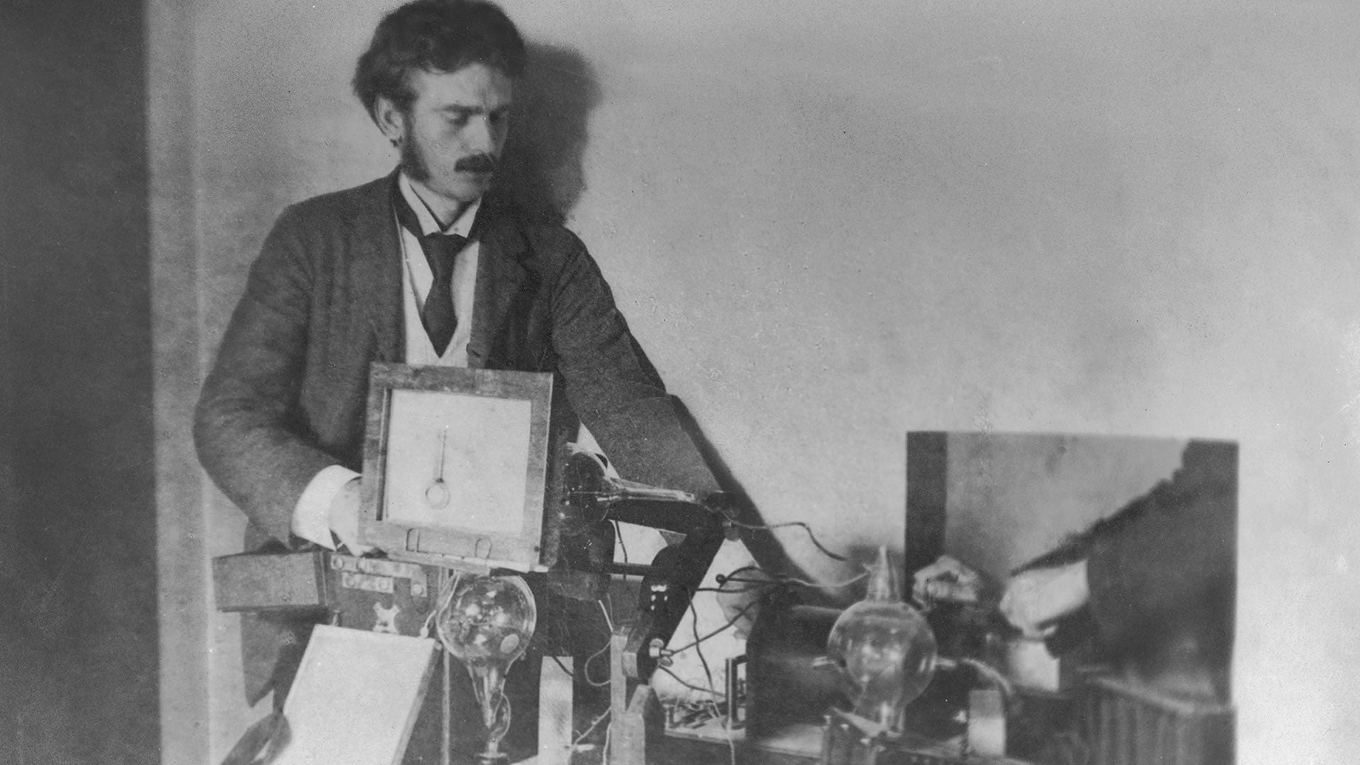
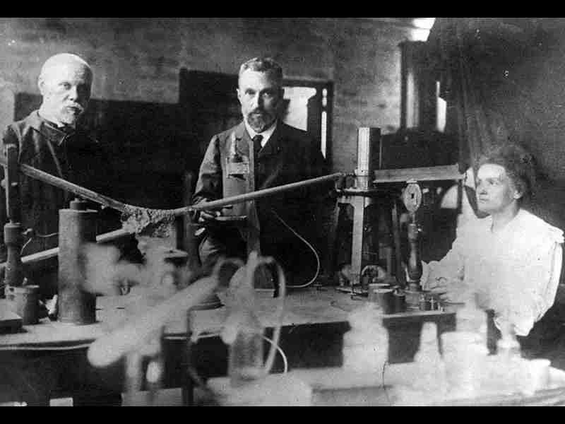
The discovery of X-rays marked a turning point in the history of science and medicine, unlocking a realm of invisible phenomena and revolutionizing our understanding of the natural world. While many individuals contributed to our understanding of X-rays, one name stands out as the pivotal figure in their discovery—Wilhelm Conrad Roentgen.

The foundations for the discovery of X-rays were laid by earlier pioneers in the late 19th century. Sir William Crookes, a British physicist, made significant strides in the study of cathode rays and their behavior within Crookes tubes. His experiments with these vacuum tubes, which contained low-pressure gases, revealed the presence of mysterious rays that emitted from the cathode and produced fluorescence on nearby materials.
Wilhelm Conrad Roentgen: The Discoverer of X-rays
In 1895, Wilhelm Conrad Roentgen, a German physicist, made the groundbreaking discovery that would propel X-rays into the realm of scientific fame. Working at the University of Würzburg, Roentgen was investigating the properties of cathode rays when he stumbled upon an unexpected phenomenon. He noticed that a fluorescent screen in his lab started to emit a peculiar glow when a nearby Crookes tube was energized, even though it was shielded from direct cathode ray exposure.
Intrigued by this phenomenon, Roentgen carefully conducted further experiments to understand the nature of these mysterious rays. He discovered that they possessed unique properties—they could pass through various materials, cast shadows, and could expose photographic plates. Recognizing the significance of his discovery, Roentgen named these new rays “X-rays,” using the mathematical symbol “X” to represent their unknown nature.
Roentgen’s discovery of X-rays sparked worldwide excitement and fascination. Scientists and physicians from around the globe rushed to explore the capabilities and potential applications of this new phenomenon. Roentgen’s work earned him the first Nobel Prize in Physics in 1901, firmly establishing him as the discoverer of X-rays.
Further Developments: The Role of Other Scientists
While Roentgen’s pivotal role in the discovery of X-rays is indisputable, subsequent researchers played significant roles in expanding our understanding and harnessing the potential of this new phenomenon.
One such scientist was Arthur Schuster, a British physicist who studied the nature of X-rays and their interaction with matter. Schuster’s experiments with X-rays led to the discovery of X-ray diffraction, a technique that would later become instrumental in the field of crystallography, enabling scientists to determine the atomic and molecular structures of various substances.
Another notable figure in X-ray research was Max von Laue, a German physicist. Building on Schuster’s work, von Laue conceived the idea that X-rays could be diffracted by crystals, forming distinctive patterns that could be analyzed to unravel the arrangement of atoms within the crystal lattice. This groundbreaking concept laid the foundation for the field of X-ray crystallography, which would later play a vital role in the discovery of the structures of DNA and proteins.
The discovery of X-rays revolutionized numerous fields, leaving an indelible impact on medicine, industry, and scientific research. In medicine, X-rays quickly became an invaluable tool for diagnostic imaging, enabling physicians to visualize internal structures, detect fractures, and diagnose diseases. The use of X-rays
History of Radiography:

Radiography, a fundamental component of medical imaging, has revolutionized healthcare by enabling the visualization of internal structures and aiding in the diagnosis of various conditions. We delve into the rich history of radiography, tracing its origins from the groundbreaking discoveries of Wilhelm Conrad Roentgen to the advancements in technology and techniques that have shaped modern medical imaging. Embark on a captivating journey through time as we explore the pioneers, key developments, and transformative impact of radiography on healthcare.
Discovery of X-rays and Early Pioneers
The story of radiography begins with the serendipitous discovery of X-rays by Wilhelm Conrad Roentgen in 1895. Roentgen, a German physicist, observed the emission of mysterious rays that could penetrate objects and create images on photographic plates. His groundbreaking work earned him the title of the discoverer of X-rays, setting the stage for the development of radiography.
Following Roentgen’s discovery, other pioneers emerged to further explore and harness the power of X-rays. Thomas Edison, renowned for his numerous inventions, contributed to the advancement of radiography by developing the first commercially available X-ray fluoroscope. Additionally, Clarence Dally and Elihu Thomson made significant contributions to X-ray tube design and electrical apparatuses, playing vital roles in the early development of radiography.
Advancements in Technology and Techniques
As radiography gained momentum, advancements in technology and techniques paved the way for more accurate and detailed imaging. One significant breakthrough was the introduction of image intensifiers in the mid-20th century, which enhanced the visibility of X-ray images, particularly in fluoroscopy. This innovation facilitated real-time imaging during procedures, allowing for dynamic visualization of anatomical structures and functions.
The advent of computed tomography (CT) in the 1970s revolutionized radiographic imaging further. The invention of the CT scanner by Godfrey Hounsfield and Allan Cormack enabled the acquisition of cross-sectional images, providing three-dimensional views of internal structures with unprecedented detail. CT scans became instrumental in diagnosing various conditions, guiding surgical interventions, and monitoring treatment responses.
Digital Radiography and Advancements in Imaging Modalities
In recent decades, the transition from analog to digital radiography has significantly enhanced imaging capabilities, efficiency, and patient care. The introduction of digital detectors, such as computed radiography (CR) and digital radiography (DR) systems, eliminated the need for traditional film processing. Digital imaging allowed for immediate image acquisition, manipulation, and sharing, reducing patient exposure to radiation and enabling efficient image storage and retrieval.
Moreover, radiography has expanded beyond conventional X-ray imaging. Modalities such as mammography, fluoroscopy, angiography, and interventional radiology have emerged, utilizing specialized techniques and equipment to visualize specific anatomical regions and perform minimally invasive procedures. These advancements have facilitated early detection of breast cancer, precise guidance during interventional procedures, and improved patient outcomes.
Radiography in Modern Healthcare
Radiography has become an integral component of modern healthcare, playing a crucial role in diagnosing and managing a wide range of medical conditions. It assists in detecting fractures, evaluating lung diseases, identifying tumors, and guiding various therapeutic interventions. Radiographic imaging, alongside other medical imaging modalities, contributes to more accurate diagnoses, enables personalized treatment plans, and improves patient outcomes.
Wilhelm Röntgen and X-ray:

The discovery of X-rays by Wilhelm Röntgen in 1895 marked a revolutionary milestone in the history of science and medicine. Röntgen’s accidental encounter with these mysterious rays unveiled a new realm of invisible phenomena, transforming our understanding of the natural world.
Wilhelm Conrad Röntgen was born on March 27, 1845, in Lennep, Prussia (now Remscheid, Germany). He pursued his education in mechanical engineering at the Polytechnic Institute in Zurich, Switzerland, where he developed a deep interest in physics. Röntgen’s passion for scientific inquiry led him to pursue further studies in physics, focusing on the properties of electricity and magnetism.
Discovery of X-rays
In 1895, while working as a professor of physics at the University of Würzburg in Germany, Röntgen made an accidental and monumental discovery. While investigating the properties of cathode rays, he noticed that a fluorescent screen in his laboratory emitted a glow when a nearby Crookes tube was energized, even though the tube was shielded from direct cathode ray exposure.
Intrigued by this unexpected phenomenon, Röntgen conducted meticulous experiments to understand its nature. He discovered that the rays possessed unique properties—they could penetrate various materials, cast shadows, and expose photographic plates. Recognizing the significance of his findings, Röntgen named these new rays “X-rays,” using the mathematical symbol “X” to represent their unknown nature.
Röntgen‘s discovery of X-rays sparked a worldwide sensation, captivating the scientific community and the public alike. His pioneering research laid the foundation for the field of radiography and revolutionized medical diagnosis and treatment. X-ray imaging, also known as roentgenography in honor of Röntgen, became an essential tool in medicine, enabling physicians to visualize internal structures, detect fractures, and diagnose diseases.
The impact of Röntgen‘s discovery extended beyond the field of medicine. X-rays found applications in diverse areas, including industry, security, and scientific research. In industry, X-ray technology facilitated non-destructive testing of materials, ensuring product quality and structural integrity. In security, X-ray scanners became crucial in detecting prohibited items in airports and other high-security areas.
Röntgen‘s pioneering work earned him numerous accolades, including the first Nobel Prize in Physics in 1901. His discovery propelled him to international fame and established him as a prominent figure in the scientific community.
The legacy of Wilhelm Röntgen and his discovery of X-rays continues to shape scientific research and medical practice. Radiology, the branch of medicine that employs X-ray imaging, has evolved with advancements in technology, including digital radiography, computed tomography (CT), mammography, and interventional radiology. These innovations have further enhanced the capabilities of medical imaging, allowing for more accurate diagnoses, less invasive procedures, and improved patient outcomes.
Charles Glover Barkla and X-ray: Unraveling the Mysteries of Radiation

The study of radiation and its properties has been a central focus of scientific inquiry for over a century. Among the notable pioneers in this field is Charles Glover Barkla, whose groundbreaking research on X-rays expanded our understanding of electromagnetic radiation and earned him the Nobel Prize in Physics in 1917.
Born on June 7, 1877, in Widnes, England, Charles Glover Barkla exhibited an early aptitude for mathematics and science. He pursued his higher education at Trinity College, Cambridge, where he studied Natural Sciences. It was during his time at Cambridge that Barkla developed a particular interest in the emerging field of X-ray research.
Research on X-rays
After completing his studies, Barkla embarked on a series of experiments to investigate the properties of X-rays, building upon the discoveries of Wilhelm Conrad Röntgen. He focused his research on the phenomenon known as X-ray scattering, in which X-rays interact with matter and change direction. Barkla meticulously studied the scattering of X-rays and the patterns they produced, seeking to unravel the underlying principles governing this phenomenon.
In 1903, Barkla made a significant breakthrough when he discovered that X-ray scattering was not uniform for all elements. He observed that different elements exhibited unique scattering patterns, leading him to propose that the scattering of X-rays was dependent on the atomic structure of the material being examined. This groundbreaking finding laid the foundation for the field of X-ray spectroscopy and opened new avenues for the study of atomic and molecular structures.
X-ray Spectroscopy and Nobel Prize
Barkla’s work on X-ray spectroscopy earned him widespread recognition. In 1917, he was awarded the Nobel Prize in Physics for his discovery of the characteristic X-ray radiation of elements and the laws governing X-ray scattering. His research provided valuable insights into the composition and arrangement of atoms within materials, shedding light on the fundamental properties of matter.
Barkla’s findings were instrumental in advancing our understanding of X-ray technology and its applications. His work led to the development of techniques such as X-ray crystallography and X-ray diffraction, which have become crucial tools in fields such as chemistry, physics, and material science.
Charles Glover Barkla’s contributions to the field of X-ray research continue to have a lasting impact. His discoveries expanded our knowledge of electromagnetic radiation, revealing the intricacies of X-ray scattering and the unique spectral signatures of different elements. Barkla’s work provided a basis for subsequent research on atomic and molecular structures, influencing advancements in spectroscopy and the understanding of chemical bonds.
Moreover, Barkla’s findings paved the way for practical applications of X-ray technology in diverse fields. X-ray imaging techniques, such as radiography and computed tomography (CT), have revolutionized medicine by enabling the non-invasive visualization of internal structures and aiding in the diagnosis of various conditions. The development of X-ray-based techniques for materials analysis, such as X-ray fluorescence and X-ray crystallography, has transformed research in fields ranging from archaeology to pharmaceuticals.
Max von Laue and X-ray:

The discovery of X-ray diffraction by Max von Laue in 1912 marked a significant milestone in the field of crystallography, unlocking the key to understanding the arrangement of atoms in crystals. Von Laue’s groundbreaking work revolutionized our understanding of the atomic world and laid the foundation for countless scientific advancements.
Max Theodor Felix von Laue was born on October 9, 1879, in Pfaffendorf, Germany. He developed a strong interest in physics from an early age and pursued his education at the University of Strasbourg and the Ludwig Maximilian University of Munich. Under the guidance of renowned scientists, including Max Planck and Arnold Sommerfeld, von Laue honed his skills and developed a deep understanding of theoretical physics.
X-ray Diffraction Experiment
Inspired by the earlier work of Charles Glover Barkla on X-ray scattering, von Laue set out to investigate whether X-rays could be diffracted by crystals. Building on the wave theory of light, von Laue hypothesized that the regular arrangement of atoms in a crystal lattice would cause X-rays to diffract, producing a distinctive pattern.
In 1912, von Laue, along with his collaborators Friedrich and Knipping, performed a groundbreaking experiment that confirmed his hypothesis. They exposed a crystal of zinc sulfide to a beam of X-rays and observed a series of dark spots on a photographic plate placed behind the crystal. These spots formed a diffraction pattern, proving that X-rays indeed undergo diffraction when passing through a crystal lattice.
Von Laue’s discovery of X-ray diffraction opened up new possibilities for studying the structure of matter. His experiment demonstrated that X-rays could be used as a powerful tool to investigate the arrangement of atoms and molecules in crystals. This breakthrough laid the foundation for the field of X-ray crystallography, which has since become an indispensable method for determining the atomic and molecular structures of a wide range of substances.
The impact of von Laue’s work reverberated across various scientific disciplines. X-ray crystallography became instrumental in elucidating the structures of complex biological molecules, such as DNA and proteins, leading to groundbreaking discoveries in genetics, biochemistry, and pharmaceutical research. The technique also found applications in materials science, solid-state physics, and chemistry, enabling scientists to understand the properties and behavior of materials at the atomic level.
Max von Laue’s contributions to science earned him numerous accolades and recognition. In 1914, he was awarded the Nobel Prize in Physics for his discovery of the diffraction of X-rays by crystals. This prestigious honor solidified von Laue’s place among the scientific greats and underscored the significance of his work.
Von Laue’s legacy extends beyond his groundbreaking experiment. He played a pivotal role in fostering international collaboration and advancing scientific education. As a professor at the University of Berlin and later the University of Göttingen, von Laue mentored a generation of scientists, including James Franck, Peter Debye, and Werner Heisenberg, who went on to make their own groundbreaking contributions to physics.
Nikola Tesla and the Discovery of X-ray Radiation:

Nikola Tesla, the visionary inventor and electrical engineer, is renowned for his groundbreaking contributions to the fields of electricity and magnetism. While Tesla is widely celebrated for his inventions such as the alternating current (AC) system and wireless transmission of energy, his involvement in the discovery of X-ray radiation remains a lesser-known aspect of his illustrious career.
Born on July 10, 1856, in Smiljan, Croatia (then part of the Austrian Empire), Tesla showed exceptional aptitude in mathematics and physics from a young age. After completing his education in engineering and physics in Graz and Prague, Tesla embarked on a career that would define him as one of the greatest inventors of his time.
Tesla’s X-ray Experiments
In the late 19th century, Nikola Tesla conducted extensive experiments in his laboratory in New York City, exploring various aspects of electrical and electromagnetic phenomena. While working with high-frequency alternating currents, Tesla observed peculiar effects that hinted at the presence of X-ray radiation.
One notable experiment involved Tesla passing high-frequency currents through a gas-filled tube. He observed that when the tube contained certain substances, it emitted a glow resembling X-rays. Tesla hypothesized that these emissions were the result of X-ray radiation being produced within the tube.
Inventors and the Discovery of X-rays
It is essential to note that Tesla‘s observations regarding X-ray radiation were made around the same time as the discoveries of Wilhelm Conrad Röntgen and Henri Becquerel. Röntgen, a German physicist, is generally credited with the discovery of X-rays in 1895. His experiments with cathode rays and their ability to penetrate matter led to the identification of this new form of electromagnetic radiation. Similarly, Becquerel, a French physicist, discovered radioactivity through his investigations into uranium salts.
While Tesla did not explicitly claim to have discovered X-rays, his experiments and observations were significant contributions to the emerging field of X-ray research. His work showcased the potential of high-frequency currents in producing X-ray-like emissions and sparked further investigations into the properties and applications of X-ray radiation.
Nikola Tesla‘s experiments with X-ray radiation laid the groundwork for future advancements in the field. His observations highlighted the potential of high-frequency currents in generating X-rays, which would later prove instrumental in medical and industrial applications. The ability of X-rays to penetrate matter and create images of internal structures revolutionized medicine, allowing for non-invasive diagnosis and treatment.
Furthermore, Tesla‘s work on X-rays influenced subsequent developments in radiography, radiotherapy, and imaging technology. The discovery of X-ray radiation opened up new frontiers in scientific research and paved the way for advancements in fields such as medical imaging, material analysis, and non-destructive testing.
Radiation Sources: Exploring the Origins and Applications of Electromagnetic Energy

Radiation sources play a vital role in a wide range of scientific, industrial, and medical applications. These sources produce electromagnetic energy, allowing us to explore the mysteries of the universe, power our modern technologies, and improve our understanding of the world around us.
Electromagnetic Spectrum: The Range of Radiation
The electromagnetic spectrum encompasses a vast range of radiation, each with its own unique properties and applications. From radio waves to gamma rays, the electromagnetic spectrum spans an incredible breadth of energy. Notable segments of the electromagnetic spectrum include radio waves, microwaves, infrared radiation, visible light, ultraviolet radiation, X-rays, and gamma rays.
Natural Sources of Radiation
Radiation can originate from both natural and artificial sources. In nature, various processes generate radiation. For example, the sun emits a vast spectrum of electromagnetic radiation, including visible light, infrared, and ultraviolet rays. Henri Becquerel discovered natural radioactivity in 1896, revealing that certain elements, such as uranium and radium, emit radiation spontaneously.
Artificial Sources of Radiation
Humans have harnessed the power of radiation for diverse applications. Wilhelm Conrad Roentgen‘s discovery of X-rays in 1895 opened up new possibilities for medical imaging and diagnostics. X-ray machines, such as X-ray tubes and CT scanners, generate X-rays for imaging bones, organs, and tissues. Nikola Tesla‘s experiments with high-frequency currents hinted at the production of X-ray-like emissions.
Another significant artificial source of radiation is radioactive isotopes. Marie Curie‘s pioneering work with radioactive elements, including radium and polonium, led to advancements in cancer treatment and the development of radiation therapy. Radioactive isotopes are used in various medical procedures, such as nuclear medicine imaging and targeted cancer treatments.
Particle Accelerators and Synchrotrons
Particle accelerators, such as the Large Hadron Collider (LHC) at CERN in Geneva, Switzerland, are powerful tools for research. These massive machines accelerate particles to high speeds, creating collisions that generate intense radiation. Particle accelerators have contributed to groundbreaking discoveries in particle physics, helping us understand the fundamental structure of matter and the origins of the universe.
Synchrotrons are a type of particle accelerator that produce high-intensity radiation called synchrotron radiation. These facilities, such as the Advanced Photon Source (APS) at Argonne National Laboratory in the United States, produce brilliant X-rays and other forms of electromagnetic radiation. Synchrotron radiation is utilized in various research fields, including materials science, chemistry, biology, and archaeology.
Radiation sources have a profound impact on numerous fields. In medicine, radiation is used for diagnostic imaging, radiotherapy, and nuclear medicine procedures. It aids in the detection and treatment of diseases such as cancer and plays a crucial role in medical research.
Industrial applications of radiation include non-destructive testing, where X-rays and gamma rays are used to inspect the integrity of materials and structures. Radiation is also utilized in sterilization processes for medical equipment and food preservation, ensuring safety and extending shelf life.
Research fields benefit greatly from radiation sources. Synchrotrons enable advanced techniques such as X-ray crystallography and spectroscopy, allowing scientists to investigate the atomic and molecular structures of materials. Radiation sources also play a crucial role in astrophysics, allowing astronomers to study celestial objects and phenomena that emit various forms of electromagnetic radiation.
Innovations in radiation sources continue to drive scientific progress. Advancements in technology have led to the development of more powerful and efficient sources, such as free-electron lasers (FELs) and plasma-based sources. FELs produce intense, coherent X-ray pulses, enabling researchers to probe matter at the atomic and molecular levels with unprecedented precision. Plasma-based sources utilize plasma physics principles to generate ultrafast and high-energy radiation pulses for a wide range of applications.
While radiation sources offer immense benefits, it is crucial to prioritize safety and adhere to strict regulations. Exposure to excessive radiation can have harmful effects on human health and the environment. Stringent measures, including radiation shielding, personal protective equipment, and dose monitoring, are implemented to ensure the safety of workers and the public in settings where radiation is used.
Government bodies and international organizations, such as the International Atomic Energy Agency (IAEA) and national radiation protection agencies, establish guidelines and regulations to ensure the safe and responsible use of radiation sources. These regulations encompass areas such as radiation safety, security, and waste management to mitigate potential risks and ensure the beneficial and ethical application of radiation technology.
X-Ray Generators: Power of Electromagnetic Imaging

X-ray generators are fundamental devices in medical and industrial imaging, enabling the creation of detailed images that reveal the internal structures of objects. These generators, pioneered by visionary inventors, produce the high-energy X-ray radiation necessary for a wide range of applications.
Early Beginnings and Wilhelm Conrad Roentgen’s Discovery
The story of X-ray generators traces back to the groundbreaking discovery of X-rays by Wilhelm Conrad Roentgen in 1895. Roentgen’s accidental discovery revealed the ability of X-rays to penetrate various materials and create images on photographic plates. This discovery laid the foundation for the development of X-ray technology and spurred the invention of X-ray generators.
Early X-ray Generators and William David Coolidge
In the early years of X-ray technology, William David Coolidge made significant contributions to the development of X-ray generators. In 1913, Coolidge invented the Coolidge X-ray tube, a crucial component in X-ray generators that produced a more stable and controllable X-ray beam. His design incorporated a heated filament to produce electrons, which were accelerated towards a target material, generating X-rays. The Coolidge X-ray tube became the standard design for X-ray generators for several decades.
Technological Advancements and High-Frequency Generators
In the early 20th century, advancements in electronics and vacuum tube technology led to the development of high-frequency X-ray generators. Valdemar Poulsen introduced the concept of high-frequency oscillators in 1903, which were later adapted for use in X-ray generators. High-frequency generators provided more efficient X-ray production and allowed for faster exposure times, improving the quality and efficiency of X-ray imaging.
Modern X-ray Generators and Digital Imaging
With the advent of digital technology, X-ray generators underwent significant advancements. The transition from analog to digital imaging brought forth digital radiography (DR) and computed radiography (CR) systems. These systems eliminated the need for film and introduced the direct capture and storage of X-ray images, enhancing image quality and facilitating faster image retrieval.
Additionally, the development of pulsed X-ray generators enabled the production of short, intense bursts of X-rays, which proved valuable in dynamic imaging techniques such as fluoroscopy and interventional radiology. Pulsed generators offered improved image clarity and reduced patient radiation exposure.
Key Contributors and Ongoing Innovations
Throughout the history of X-ray generators, numerous inventors and researchers have contributed to their development. Besides Wilhelm Conrad Roentgen, William David Coolidge, and Valdemar Poulsen, other notable names include Peter Guthrie Tait, Kenneth John Button, and Arthur Holly Compton. These individuals played crucial roles in advancing the technology and applications of X-ray generators.
Today, ongoing innovations in X-ray generator technology focus on improving image resolution, reducing radiation dose, and enhancing workflow efficiency. These advancements include the development of digital detectors, low-dose imaging techniques, multi-energy imaging, and mobile X-ray systems. These innovations continue to push the boundaries of X-ray imaging, enabling more precise diagnoses, minimally invasive procedures, and improved patient care.
Present State of Radiography:

Radiography, a fundamental pillar of medical imaging, has evolved significantly since its inception. From the pioneering work of Wilhelm Conrad Roentgen to the present day, radiography has undergone remarkable advancements in technology, applications, and patient care.
The birth of radiography can be traced back to the discovery of X-rays by Wilhelm Conrad Roentgen in 1895. Roentgen’s accidental discovery led to the rapid development of X-ray technology and the subsequent invention of the X-ray machine. Roentgen’s work earned him the first Nobel Prize in Physics in 1901 and laid the foundation for the field of radiography.
Advancements in technology have transformed radiography, particularly with the digital revolution. Traditional film-based radiography has largely been replaced by digital radiography (DR) and computed radiography (CR) systems. These digital imaging modalities offer several advantages, including improved image quality, reduced radiation exposure, and enhanced workflow efficiency. The shift to digital imaging has revolutionized the way radiographic images are acquired, stored, and shared.
Computed Tomography (CT) and Three-Dimensional Imaging
The advent of computed tomography (CT) has revolutionized diagnostic imaging. CT scanners utilize X-rays to create cross-sectional images of the body, providing detailed insights into anatomical structures and pathology. The ability to reconstruct three-dimensional images from CT data has further enhanced diagnostic accuracy and treatment planning.
Magnetic Resonance Imaging (MRI) and Nuclear Medicine
Radiography extends beyond X-rays. Magnetic Resonance Imaging (MRI) utilizes powerful magnets and radio waves to produce highly detailed images of soft tissues and organs. MRI has become a valuable tool in diagnosing a wide range of conditions, from brain disorders to musculoskeletal injuries. Additionally, nuclear medicine techniques, such as positron emission tomography (PET) and single-photon emission computed tomography (SPECT), combine radiopharmaceuticals and imaging to visualize physiological processes at the molecular level.
Interventional Radiology and Image-Guided Procedures
Interventional radiology has revolutionized minimally invasive procedures. Using real-time imaging guidance, interventional radiologists can perform procedures such as angioplasty, stent placement, and tumor ablation with precision and minimal invasiveness. These techniques reduce the need for open surgery, resulting in shorter hospital stays, faster recovery times, and improved patient outcomes.
Radiation Safety and Dose Reduction
In recent years, significant emphasis has been placed on radiation safety and dose reduction in radiography. Stricter regulations, advanced shielding techniques, and dose monitoring systems ensure the safety of patients and healthcare professionals. Technology advancements, such as low-dose imaging protocols and optimization algorithms, further minimize radiation exposure while maintaining image quality, making radiography safer than ever.
Artificial Intelligence (AI) and Machine Learning
The integration of artificial intelligence (AI) and machine learning algorithms has the potential to revolutionize radiography. AI applications can assist radiologists in image interpretation, workflow optimization, and detection of abnormalities. Deep learning algorithms can analyze vast amounts of radiographic data and provide accurate and efficient diagnoses, improving the overall efficiency and accuracy of radiographic interpretations.
Key Dates at X-ray Discovery:

The discovery of X-rays by Wilhelm Conrad Roentgen in 1895 opened up a new era in medical imaging and scientific exploration. From the initial discovery to advancements in technology and applications, we explore the chronological milestones that have revolutionized the field of X-rays.
1895: Wilhelm Conrad Roentgen and the Discovery of X-rays
On November 8, 1895, Wilhelm Conrad Roentgen, a German physicist, made a remarkable discovery while experimenting with cathode rays. Working in his laboratory at the University of Würzburg, Roentgen noticed a fluorescent glow emitted by a barium platinocyanide screen placed nearby. He realized that this glow was caused by a new and mysterious form of radiation. Naming it “X-rays” due to their enigmatic nature, Roentgen published his findings in a seminal paper titled “On a New Kind of Rays” and revolutionized the field of medical imaging.
1896: Henri Becquerel and the Discovery of Radioactivity
In 1896, French physicist Henri Becquerel made a significant contribution to the understanding of X-rays. While studying the properties of uranium salts, Becquerel accidentally discovered radioactivity. He found that uranium emitted a type of radiation similar in penetrating power to X-rays. This discovery paved the way for further investigations into radioactive elements and their role in emitting radiation.
Early 20th Century: Medical Applications of X-rays
The early 20th century witnessed the rapid development of X-ray applications in medicine. In 1901, British surgeon John Macintyre became one of the first physicians to utilize X-rays for medical purposes. He successfully visualized a bullet lodged in a patient’s wrist using X-ray imaging. This marked the beginning of X-ray diagnostics in surgical procedures.
1913: William David Coolidge and the Coolidge X-ray Tube
In 1913, American physicist William David Coolidge revolutionized X-ray technology with his invention of the Coolidge X-ray tube. This innovative device utilized a heated tungsten filament to produce a more stable and controllable X-ray beam. The Coolidge X-ray tube superseded earlier designs, offering improved reliability, higher-quality X-ray images, and enhanced safety for both patients and operators.
1963: Godfrey Hounsfield and the Invention of Computed Tomography (CT)
In 1963, British engineer Godfrey Hounsfield introduced a groundbreaking technology that would transform medical imaging: computed tomography (CT). Hounsfield’s invention combined X-ray imaging with computer algorithms to generate detailed cross-sectional images of the body. CT scans provided physicians with unprecedented insights into anatomical structures, greatly enhancing diagnostic capabilities.
Late 20th Century: Digital Radiography and Imaging Advancements
In the late 20th century, digital imaging revolutionized the field of radiography. Pioneers such as Steven M. Horii and Eliot L. Siegel made significant contributions to the development of digital radiography (DR), which replaced traditional film-based systems with digital detectors. This transition allowed for instant image acquisition, storage, and manipulation, improving workflow efficiency and image quality.
1996: Introduction of Digital Subtraction Angiography (DSA)
In 1996, the introduction of digital subtraction angiography (DSA) further advanced X-ray imaging capabilities. DSA utilized digital imaging techniques to selectively
subtract the non-vascular structures from X-ray images, enhancing the visualization of blood vessels. This technique revolutionized the field of interventional radiology, enabling precise diagnosis and treatment of vascular conditions.
21st Century: Advancements in X-ray Technology
The 21st century has seen remarkable advancements in X-ray technology, driving further innovation and improved patient care. Key developments include:
– Cone Beam CT (CBCT): Cone beam CT imaging, also known as digital volume tomography, offers high-resolution three-dimensional imaging with a cone-shaped X-ray beam. It has become particularly valuable in dental and maxillofacial imaging, providing detailed views of the teeth, jaws, and facial structures.
– Digital Breast Tomosynthesis (DBT): Digital breast tomosynthesis, or 3D mammography, has revolutionized breast cancer screening. This technique captures multiple X-ray images of the breast from different angles, producing three-dimensional reconstructions that enhance the detection of breast abnormalities.
– Dual-Energy X-ray Absorptiometry (DXA): DXA scanning is a precise method for assessing bone mineral density and diagnosing conditions such as osteoporosis. By utilizing two different X-ray energy levels, DXA scanners provide accurate measurements of bone density and aid in the evaluation of fracture risk.
– X-ray Guided Radiation Therapy (XRT): X-ray guided radiation therapy utilizes X-ray imaging during radiation treatment to precisely target tumors and spare healthy surrounding tissues. This technique ensures accurate and effective delivery of radiation, optimizing cancer treatment outcomes.
Future Directions: Artificial Intelligence (AI) and X-ray Imaging
The future of X-ray imaging holds immense promise, particularly with the integration of artificial intelligence (AI). AI algorithms have the potential to enhance image interpretation, improve workflow efficiency, and aid in the early detection of abnormalities. Machine learning techniques can analyze vast amounts of X-ray data, assisting radiologists in making accurate diagnoses and providing personalized patient care.
Conclusion
In conclusion, the discovery and invention of X-rays have revolutionized the field of medical imaging and had a profound impact on various scientific disciplines. The question of “Who invented X-ray?” has a definitive answer: it was Wilhelm Conrad Roentgen, a German physicist, who made the groundbreaking discovery in 1895. Roentgen’s accidental finding of X-rays, their ability to penetrate matter, and their unique properties opened up new possibilities for exploring the human body and understanding the fundamental nature of radiation.
Roentgen’s work not only earned him the first Nobel Prize in Physics in 1901 but also laid the foundation for the development of radiography and radiology as vital components of modern healthcare. However, it is essential to acknowledge that the discovery of X-rays was built upon the collective knowledge and contributions of several scientists who preceded Roentgen.
Scientists such as Henri Becquerel and William David Coolidge played significant roles in advancing our understanding and application of X-rays. Becquerel’s discovery of radioactivity in 1896 and Coolidge’s invention of the Coolidge X-ray tube in 1913 were instrumental in furthering the field. Their contributions enhanced the safety, reliability, and quality of X-ray technology, making it an invaluable tool for medical diagnostics, industrial applications, and scientific research.
Throughout the history of X-rays, numerous inventors, researchers, and pioneers have contributed to the advancement of this field. Names such as John Macintyre, Francis Williams, Godfrey Hounsfield, Steven M. Horii, and Eliot L. Siegel have played crucial roles in developing new techniques, imaging modalities, and digital technologies that have transformed X-ray imaging.
The invention of X-rays has had a profound impact on medical diagnosis, treatment, and research. It has revolutionized fields such as radiology, oncology, cardiology, and orthopedics, enabling early disease detection, precise interventions, and improved patient outcomes. The continuous advancements in X-ray technology, including digital imaging, computed tomography (CT), and artificial intelligence (AI), hold promise for further enhancing the accuracy, efficiency, and safety of X-ray diagnostics.
In conclusion, the invention of X-rays by Wilhelm Conrad Roentgen marked a pivotal moment in the history of science and medicine. It is through the collaborative efforts of numerous scientists and inventors that X-ray technology has evolved and continues to transform the field of medical imaging, benefiting millions of patients worldwide.
References:
- Becquerel, H. (1896). “Sur les radiations émises par phosphorescence.” Comptes Rendus de l’Académie des Sciences, 122, 420-421.
- Coolidge, W. D. (1914). “A Powerful Roentgen Ray Bulb.” Physical Review, 3(6), 313-314.
- Hounsfield, G. N. (1973). “Computerized transverse axial scanning (tomography): Part 1. Description of system.” British Journal of Radiology, 46(552), 1016-1022.
- Horii, S. M., & Siegel, E. L. (2004). “PACS and imaging informatics: Basic principles and applications.” Wiley-Blackwell.
- Macintyre, J. (1896). “The Radiography of Foreign Bodies.” The Lancet, 147(3781), 8-10.
- Roentgen, W. C. (1895). “On a New Kind of Rays.” Nobel Lecture, December 11, 1901.
- Siegel, E. L., Reiner, B. I., & Carrino, J. A. (2012). “Medical Imaging Informatics.” Springer Science & Business Media.
- Williams, F. J. (1901). “On the penetration of glass and other matters by Röntgen rays.” Philosophical Magazine, 6(41), 573-586.
- Macovski, A. (1983). “Medical Imaging Systems.” Prentice-Hall.
- Bushberg, J. T., Seibert, J. A., Leidholdt Jr, E. M., & Boone, J. M. (2011). “The Essential Physics of Medical Imaging.” Lippincott Williams & Wilkins.
- Halpern, E. J., & D’Orsi, C. J. (2012). “Breast Imaging.” Elsevier Health Sciences.
- Bushong, S. C. (2018). “Radiologic Science for Technologists: Physics, Biology, and Protection.” Elsevier Health Sciences.
- McNitt-Gray, M. F. (2009). “AAPM/RSNA physics tutorial for residents: topics in CT: radiation dose in CT.” Radiographics, 29(2), 517-531.
- Lang, N., & Efremidis, K. (2017). “Emerging Techniques in Diagnostic Radiology: Multidetector-Row CT.” Springer.
- Thrall, J. H. (2013). “Radiology Review Manual.” Lippincott Williams & Wilkins.


