The discovery of X-rays revolutionized the field of medical imaging and profoundly impacted various scientific disciplines. The question of “who invented X-ray?” leads us to the remarkable individuals whose ingenuity and perseverance paved the way for this groundbreaking technology. In this article, we delve into the key inventors and pioneers who played a significant role in the development of X-rays, uncovering their contributions and the impact they had on the field of radiography.

Wilhelm Conrad Roentgen: The Discoverer of X-rays
The journey of X-rays began with the groundbreaking work of Wilhelm Conrad Roentgen. In 1895, while conducting experiments on cathode rays, Roentgen made an extraordinary observation. He noticed that a fluorescent screen in his lab glowed even though it was not in direct contact with the cathode rays. Intrigued by this phenomenon, Roentgen conducted further experiments, eventually realizing that he had discovered an entirely new form of radiation – X-rays.
Roentgen’s pivotal discovery earned him the first Nobel Prize in Physics in 1901. His work laid the foundation for the field of radiography, and the term “X-ray” was coined in his honor. Roentgen’s exploration of X-rays opened up vast possibilities for medical diagnosis and scientific investigation.
Henri Becquerel: The Pioneer of Radioactivity
While Roentgen’s discovery of X-rays was a breakthrough, it was Henri Becquerel who further expanded our understanding of radiation. In 1896, Becquerel accidentally discovered radioactivity while studying the properties of uranium salts. He found that these substances emitted a type of radiation that could penetrate opaque materials, similar to X-rays.
Becquerel’s research on radioactivity, alongside the work of Marie Curie, laid the foundation for the field of nuclear physics. Although X-rays and radioactivity are distinct phenomena, their discovery and subsequent exploration shared commonalities, contributing to our understanding of the behavior of electromagnetic radiation.
William Coolidge: The Inventor of the Modern X-ray Tube
While Roentgen and Becquerel were pivotal in the discovery and early exploration of X-rays, it was William Coolidge who made significant advancements in X-ray technology. In the early 20th century, Coolidge invented the modern X-ray tube, a device that produced a steady and controllable stream of X-rays.
Coolidge’s X-ray tube, known as the “Coolidge tube,” utilized a heated filament to emit electrons, which then struck a target material, producing X-rays. His innovation vastly improved the quality and reliability of X-ray imaging, making it an indispensable tool in medical diagnostics.
Contributions and Impact
The combined efforts of Roentgen, Becquerel, Coolidge, and numerous other researchers and inventors shaped the field of radiography and the broader domain of medical imaging. Their contributions enabled the visualization of internal structures in the human body, revolutionizing medical diagnostics and treatment.
X-rays became an essential tool in identifying fractures, tumors, and various other conditions that were previously undetectable without invasive procedures. The non-invasive nature of X-rays, coupled with their ability to provide detailed images, led to significant advancements in medical care and patient outcomes.
Moreover, X-rays found applications in fields beyond medicine. They became crucial in materials testing, security screening, and industrial inspections. X-ray technology continues to evolve, with advancements such as computed tomography (CT) scans, digital X-ray systems, and fluoroscopy techniques pushing the boundaries of diagnostic capabilities.
Brief History of Radiology

The history of radiology is a fascinating journey that has revolutionized the field of medicine. From the discovery of X-rays to the advent of advanced imaging technologies, this article explores the key milestones, inventors, and breakthroughs that have shaped the field of radiology. Tracing the evolution of medical imaging unveils a rich tapestry of innovation and scientific discovery that has forever transformed healthcare.
The Discovery of X-Rays: Wilhelm Conrad Roentgen
The history of radiology begins with the groundbreaking discovery of X-rays by Wilhelm Conrad Roentgen in 1895. While conducting experiments with cathode rays, Roentgen noticed that a fluorescent screen in his lab emitted a mysterious form of radiation. This discovery led to the birth of a new field: radiology.
Roentgen’s meticulous experiments and subsequent documentation of X-rays earned him the first Nobel Prize in Physics in 1901. His work laid the foundation for the use of X-rays in medical diagnostics and set the stage for future advancements in radiology.
Early Imaging Techniques: Radiography and Fluoroscopy
Following Roentgen’s discovery, the field of radiology rapidly advanced. In the early 20th century, radiography emerged as the primary imaging technique. Radiography involved passing X-rays through the body onto a photographic plate, creating shadow-like images that revealed anatomical structures.
In 1896, Thomas Edison and Thomas Edison Dally introduced fluoroscopy, a technique that allowed real-time X-ray visualization. Fluoroscopy involved the use of a fluorescent screen and a fluorescent material that emitted visible light when exposed to X-rays. This technique enabled dynamic imaging of structures such as the heart and gastrointestinal tract.
Radioactive Substances and Nuclear Medicine
In the early 20th century, scientists began to explore the medical applications of radioactive substances. Marie Curie and her husband, Pierre Curie, made significant contributions to this field. They discovered new radioactive elements like radium and polonium, leading to the development of nuclear medicine.
Nuclear medicine involves the use of radioactive isotopes for diagnostic and therapeutic purposes. It utilizes imaging techniques such as gamma cameras and positron emission tomography (PET) to detect and visualize radioactive tracers in the body. Nuclear medicine plays a crucial role in diagnosing and treating various diseases, including cancer and cardiovascular conditions.
Advancements in Imaging Technology: CT, MRI, and Beyond
The latter half of the 20th century witnessed groundbreaking advancements in imaging technology, expanding the capabilities of radiology. In the 1970s, the development of computed tomography (CT) scanners revolutionized diagnostic imaging. CT scanners produced cross-sectional images of the body, offering detailed information about internal structures.
In the 1980s, magnetic resonance imaging (MRI) emerged as another transformative imaging modality. MRI utilizes strong magnetic fields and radio waves to generate highly detailed images of the body’s soft tissues. It provides excellent contrast resolution and has become indispensable in diagnosing neurological disorders, musculoskeletal conditions, and more.
Furthermore, advancements in imaging technology have led to the development of digital radiography, ultrasound, and interventional radiology techniques. Digital radiography has replaced traditional film-based X-rays with digital detectors, allowing for enhanced image manipulation and storage. Ultrasound uses sound waves to create real-time images, while interventional radiology involves minimally invasive procedures guided by imaging technology.
Wilhelm Conrad Röntgen: The Discoverer of X-Rays

Wilhelm Conrad Röntgen, a renowned German physicist, is celebrated as the discoverer of X-rays, a revolutionary breakthrough in the field of science and medicine. Röntgen’s remarkable discovery not only earned him accolades but also laid the foundation for the field of radiology. This article delves into the life, achievements, and enduring legacy of Wilhelm Conrad Röntgen, highlighting his significant contributions to the scientific community.
Early Life and Education. Born on March 27, 1845, in Lennep (now Remscheid), Germany, Wilhelm Conrad Röntgen displayed an early interest in physics and engineering. He pursued his higher education at the Federal Polytechnic Institute in Zurich, Switzerland, studying mechanical engineering and graduating with distinction.
Discovery of X-Rays. Röntgen’s groundbreaking discovery of X-rays occurred on November 8, 1895, while he was conducting experiments with cathode rays. He noticed that a fluorescent screen in his laboratory emitted a mysterious glow even when shielded from direct cathode rays. Intrigued by this phenomenon, Röntgen meticulously studied and documented these rays, eventually naming them “X-rays” due to their unknown nature.
Röntgen’s experiments with X-rays revealed their remarkable ability to penetrate various materials, including human flesh, while leaving distinct shadows on photographic plates. This discovery opened up new possibilities for medical diagnostics, allowing doctors to visualize the internal structures of the human body non-invasively.
The Impact of Röntgen’s Discovery. Wilhelm Conrad Röntgen’s discovery of X-rays had an immediate and profound impact on the scientific and medical communities. His seminal paper, “On A New Kind of Rays,” published in 1895, detailed his findings and sparked worldwide interest.
Röntgen’s X-rays revolutionized the field of medicine, providing physicians with a powerful diagnostic tool. X-rays enabled the detection of fractures, tumors, and foreign bodies within the human body, offering invaluable insights for medical diagnoses. This non-invasive imaging technique quickly became an essential component of modern medical practice.
Recognition and Legacy. In recognition of his monumental achievement, Wilhelm Conrad Röntgen was awarded the first Nobel Prize in Physics in 1901. The prize acknowledged his extraordinary discovery and its significant contributions to scientific knowledge and medical advancements.
Röntgen’s discovery of X-rays laid the foundation for the field of radiology, which continues to evolve and transform healthcare. His groundbreaking work not only influenced the medical profession but also inspired further scientific exploration and technological developments in the field of imaging.
A Second Source of Radiation:

The discovery of A Second Source of Radiation represents a significant advancement in the field of physics and radiology. This groundbreaking revelation, which emerged as a complement to X-rays, shed light on a new source of radiation with unique properties and implications. In this article, we explore the origins, key inventors, and the implications of this second source of radiation, uncovering its impact on scientific understanding and applications.
Discovery of the Second Source of Radiation
The discovery of the Second Source of Radiation occurred through the diligent efforts of various researchers and inventors in the early 20th century. It emerged as a distinct entity alongside the pioneering work of Wilhelm Conrad Röntgen and his discovery of X-rays. As scientists delved deeper into the nature of radiation, they began to observe an additional source that exhibited different characteristics.
Key Inventors and Researchers
Several prominent figures played crucial roles in the identification and exploration of this second source of radiation. Notable names among these pioneers include Marie Curie, Henri Becquerel, and Ernest Rutherford. Their groundbreaking research and discoveries added layers to our understanding of radiation and its various forms.
Marie Curie and Radioactivity
Marie Curie’s work on radioactivity was instrumental in shedding light on the second source of radiation. Alongside her husband, Pierre Curie, she discovered new radioactive elements such as polonium and radium. Marie Curie’s tireless efforts and groundbreaking research earned her two Nobel Prizes—one in Physics and another in Chemistry—and cemented her status as a pioneer in the field.
Henri Becquerel and Uranium Rays
Henri Becquerel’s accidental discovery of uranium rays further contributed to the understanding of the second source of radiation. While investigating the properties of uranium salts, Becquerel noticed that these substances emitted a type of radiation that could penetrate opaque materials. This finding served as a catalyst for subsequent research on this mysterious radiation source.
Ernest Rutherford and Alpha and Beta Particles
Ernest Rutherford’s experiments with radioactive materials, particularly his investigations into alpha and beta particles, added significant insights into the second source of radiation. Rutherford’s work helped classify the different types of radiation and elucidated their distinctive properties, enabling a more comprehensive understanding of the phenomenon.
Implications and Applications
The discovery of the second source of radiation had far-reaching implications across various scientific disciplines and applications. It laid the groundwork for advancements in fields such as nuclear physics, medicine, and industry.
In the field of medicine, the second source of radiation found practical applications in diagnostic imaging, cancer treatment, and sterilization procedures. It enabled the development of techniques such as radiation therapy and radiography, further enhancing medical care and interventions.
In the realm of industry, the second source of radiation became instrumental in various fields. It facilitated the measurement and analysis of materials through techniques such as radiography testing and nuclear gauges. Additionally, it provided valuable insights into the structure of matter, atomic interactions, and the behavior of particles.
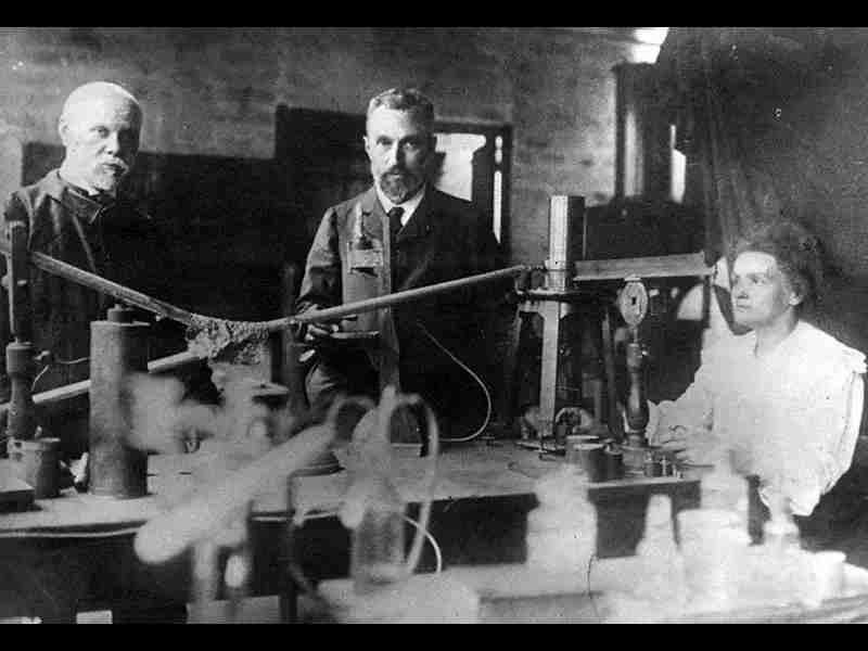
Marie Curie: Pioneering Work on Radiation and Radioactivity

Marie Curie, a remarkable scientist of the late 19th and early 20th centuries, made groundbreaking contributions to the field of radiation and radioactivity. Her relentless pursuit of knowledge and her pioneering research revolutionized our understanding of this mysterious phenomenon. This article explores Marie Curie’s work on radiation, her discoveries, and the lasting impact she had on the scientific community.
Early Life and Education. Born Maria Skłodowska on November 7, 1867, in Warsaw, Poland, Marie Curie developed an early passion for science. She pursued her education at the Sorbonne in Paris, where she met her future husband, Pierre Curie. Together, they embarked on a scientific journey that would shape the field of radiation.
Discovering Radioactivity. Marie Curie’s pivotal contribution to science began with her research on radioactivity. In the late 19th century, she and Pierre Curie investigated the properties of uranium, a radioactive element. Their experiments led to the discovery of two new elements: polonium (named after Marie’s native Poland) and radium.
Marie Curie coined the term “radioactivity” to describe the spontaneous emission of radiation from certain elements. Her work revealed that these radioactive elements emitted different forms of radiation, including alpha particles, beta particles, and gamma rays. This groundbreaking discovery challenged the prevailing understanding of matter and energy.
Isolating Radium. Marie Curie’s most notable achievement was the isolation of radium from pitchblende, a uranium-rich ore. Through meticulous chemical processes, she and Pierre Curie were able to extract minute amounts of radium. This accomplishment required tremendous dedication and perseverance, as the isolation process was arduous and dangerous due to the intense radioactivity of the element.
Recognition and Legacy. Marie Curie’s groundbreaking work on radiation earned her numerous accolades and honors. In 1903, she became the first woman to win a Nobel Prize, jointly awarded with Pierre Curie and Henri Becquerel for their research on radioactivity. In 1911, she received her second Nobel Prize, this time in Chemistry, becoming the first person and the only woman to win Nobel Prizes in two different scientific fields.
Marie Curie’s contributions to science laid the foundation for advancements in nuclear physics, medical diagnostics, and cancer treatment. Her discoveries led to the development of radiation therapy and radiography, revolutionizing medical care and saving countless lives. Her dedication and perseverance in the face of adversity continue to inspire scientists and women in STEM fields worldwide.
Tesla’s Work on Radiology:

Nikola Tesla, the visionary inventor and engineer, is widely known for his pioneering work in electrical engineering. However, his contributions to the field of radiology are often overlooked. Tesla’s unique insights and inventions in electromagnetic science laid the groundwork for advancements in radiology and wireless communication. This article explores Tesla’s work on radiology, his inventions, and the impact he had on the field.
Early Life and Background. Nikola Tesla was born on July 10, 1856, in Smiljan, Croatia. He showed an exceptional aptitude for electrical and mechanical engineering from a young age. Tesla’s passion for innovation and his understanding of electromagnetic principles became the driving force behind his revolutionary work.
Wireless Power Transmission and Radiology. Tesla’s visionary ideas and experiments paved the way for the development of wireless power transmission and its applications in radiology. His experiments with alternating current (AC), coupled with his invention of the Tesla coil, demonstrated the possibility of wireless energy transfer over long distances. This breakthrough would later influence the field of radiology.
Tesla’s Coil and High-Frequency Oscillators. One of Tesla’s most notable inventions, the Tesla coil, became an integral part of his work in radiology. This device allowed for the generation of high-voltage, high-frequency electricity, enabling experiments with electromagnetic waves and their potential applications. Tesla’s high-frequency oscillators served as precursors to the modern technology used in radiology and wireless communication.
Tesla’s Experiments with X-Rays. While Tesla is primarily associated with his work on alternating current, he also made significant contributions to the understanding and exploration of X-rays. He conducted experiments to improve the efficiency of X-ray production, focusing on generating more intense and controllable X-ray beams. Tesla’s experiments laid the foundation for advancements in X-ray technology.
Tesla’s Wireless Telegraphy and Communication. Tesla’s work on wireless power transmission not only impacted radiology but also revolutionized wireless telegraphy and communication systems. His concept of a World Wireless System aimed to transmit not only power but also information wirelessly across great distances. Tesla’s experiments and inventions, such as the Tesla oscillator and the Tesla tower, played a crucial role in the development of wireless communication technologies.
The Tesla Wardenclyffe Tower. One of Tesla’s most ambitious projects was the Wardenclyffe Tower, also known as the Tesla Tower. This monumental structure was intended to be a wireless power and communication station capable of transmitting electricity and information wirelessly around the globe. Although the Wardenclyffe Tower was never fully realized, its design and concept demonstrated Tesla’s visionary ideas for global wireless communication.
Legacy and Impact. Tesla’s work on radiology and wireless technology had a profound impact on the scientific and technological landscape. His inventions and discoveries laid the foundation for the development of modern radiology, wireless communication, and electrical engineering.
Tesla’s visionary ideas and contributions to radiology continue to shape the field. His innovations in high-frequency oscillators, wireless power transmission, and X-ray technology set the stage for advancements in medical diagnostics, radiotherapy, and telecommunications. Tesla’s pioneering work on radiology and electromagnetic science stands as a testament to his remarkable intellect and his unwavering dedication to pushing the boundaries of scientific understanding.
How Do X-Rays Work:

X-rays have become an indispensable tool in modern medicine, enabling the visualization of internal structures in the human body. But have you ever wondered how X-rays actually work? In this article, we explore the science behind X-rays, from their discovery to their application in medical diagnostics. We delve into the inventors, key concepts, and the imaging process, shedding light on the fascinating world of X-ray technology.
The Discovery of X-Rays: Wilhelm Conrad Röntgen
The story of how X-rays work begins with the pioneering work of Wilhelm Conrad Röntgen in the late 19th century. In 1895, Röntgen discovered X-rays while conducting experiments with cathode rays. He noticed that a fluorescent screen in his laboratory emitted a glow when exposed to these mysterious rays.
Understanding X-Rays: Electromagnetic Radiation
To comprehend how X-rays work, we must first understand that X-rays are a form of electromagnetic radiation. Electromagnetic radiation consists of energy waves that propagate through space. X-rays have shorter wavelengths and higher energies than visible light, making them capable of penetrating objects, including body tissues.
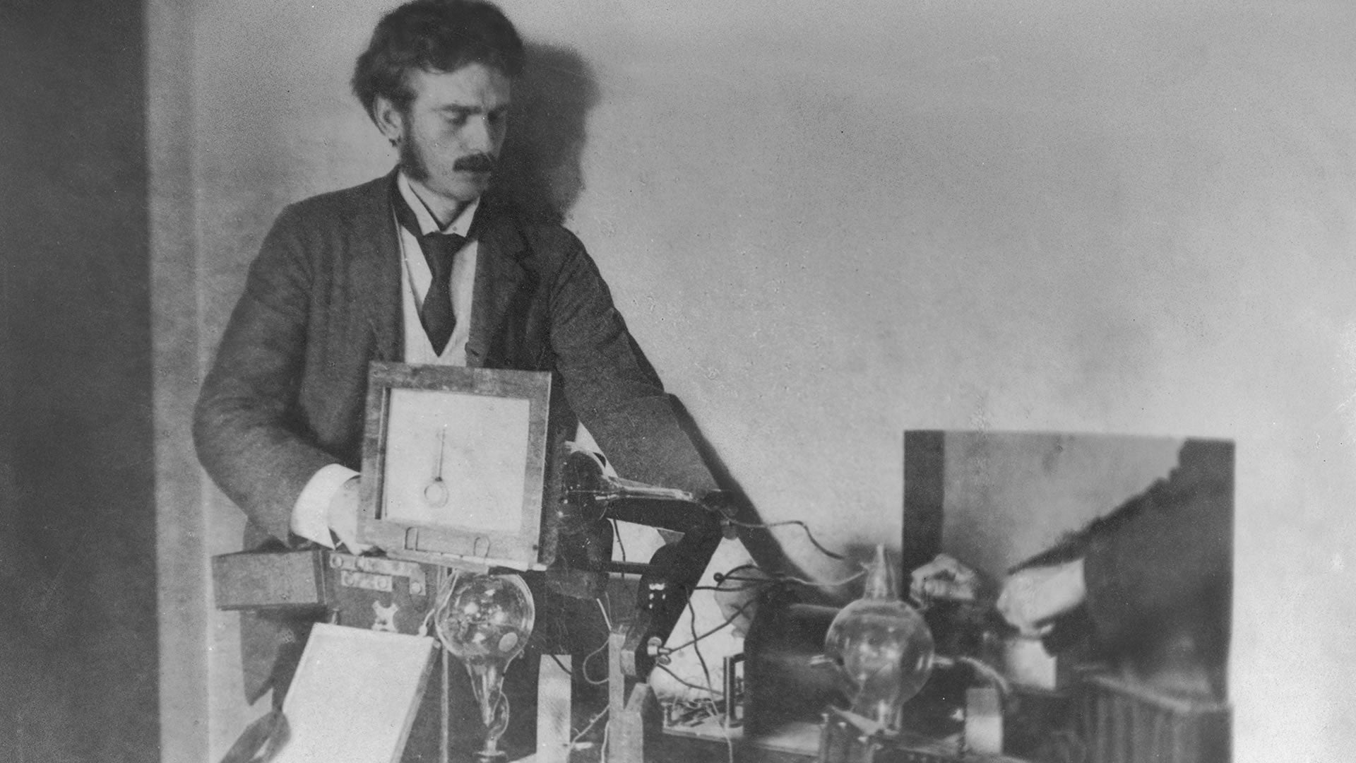
X-Ray Production: The X-Ray Tube
The primary tool used to generate X-rays is the X-ray tube. The X-ray tube consists of a cathode and an anode. When a high voltage is applied across the tube, it accelerates electrons from the cathode towards the anode. As the high-speed electrons strike the anode, they produce X-ray photons through a process called bremsstrahlung (braking radiation) and characteristic radiation.
Bremsstrahlung and Characteristic Radiation
In bremsstrahlung, electrons undergo rapid deceleration when interacting with the anode’s atomic nuclei. This deceleration results in the emission of X-ray photons with a continuous spectrum of energies. On the other hand, characteristic radiation occurs when high-speed electrons collide with inner-shell electrons of the anode material, causing the displacement of those electrons. As the displaced electrons are replaced by outer-shell electrons, X-ray photons of specific energies are emitted.
X-Ray Interactions: Absorption and Scattering
When X-rays pass through matter, they interact with the atoms in the material. These interactions can result in either absorption or scattering of the X-ray photons. Absorption occurs when X-ray photons transfer their energy to the atoms, causing ionization or excitation. Scattering, on the other hand, involves the deflection of X-ray photons without energy transfer.
X-Ray Imaging: Radiography and Computed Tomography (CT)
Now that we understand how X-rays are generated and interact with matter, let’s explore how they are used in medical imaging. The most common form of X-ray imaging is radiography. During radiography, X-rays pass through the body and interact with different tissues, resulting in varying levels of absorption. The remaining X-ray photons strike a detector, forming an image that reveals the internal structures of the body.
In more complex cases, computed tomography (CT) scans are used. CT scans involve a rotating X-ray source and detectors that create detailed cross-sectional images of the body. CT scans provide three-dimensional information and are particularly useful for diagnosing complex conditions.
X-Ray Health Concerns: Understanding the Risks and Safety Measures

X-rays have revolutionized medical diagnostics, allowing physicians to visualize internal structures and aid in the diagnosis of various conditions. While X-rays are generally considered safe and effective, it is essential to understand the potential health concerns associated with their use. In this article, we explore the risks, safety measures, and ongoing efforts to minimize radiation exposure in X-ray imaging.
Radiation Basics: Ionizing Radiation and X-Rays
X-rays are a form of ionizing radiation—radiation that carries enough energy to remove tightly bound electrons from atoms, potentially causing damage to living tissues. X-rays have higher energy levels than non-ionizing radiation, such as visible light or radio waves, making them more capable of penetrating body tissues.
Potential Health Risks: Ionizing Radiation Effects
Exposure to ionizing radiation, including X-rays, carries certain health risks. The primary concern is the potential for radiation-induced cancer. Prolonged or high-dose exposure to X-rays may increase the risk of developing certain types of cancer, particularly in organs directly exposed to the radiation.
Additionally, excessive exposure to X-rays can cause radiation burns or radiation dermatitis—skin damage resulting from high-energy X-rays damaging skin cells. It is important to note that the risk of developing health issues from X-rays is generally low when appropriate safety measures are in place.
Radiation Dose and Safety Measures
To ensure the safe use of X-rays, medical professionals adhere to strict radiation safety measures. These measures include:
- Justification: Medical imaging using X-rays is only performed when the benefits outweigh the potential risks, ensuring that the procedure is necessary for diagnosis or treatment.
- Optimization: Techniques and equipment are optimized to deliver the lowest possible radiation dose while maintaining image quality. This involves adjusting X-ray machine settings and using appropriate shielding.
- Radiation Shielding: Lead aprons, collimators, and other protective measures are used to shield areas of the body not being imaged, minimizing radiation exposure to non-target tissues.
- Radiation Monitoring: Medical professionals and technicians wear dosimeters to measure their radiation exposure levels and ensure they stay within acceptable limits.
- Pregnancy Considerations: Special care is taken for pregnant patients to minimize fetal exposure, using shielding and alternative imaging techniques when possible.
Patient Education and Informed Consent
To ensure patient safety, it is crucial for healthcare providers to educate and inform patients about the risks and benefits of X-ray procedures. This includes discussing the necessity of the procedure, potential risks, and alternative imaging options when appropriate. Informed consent allows patients to make well-informed decisions regarding their healthcare, taking into account their individual circumstances.
Ongoing Research and Technological Advancements
The field of radiology continually strives to improve patient safety and minimize radiation exposure. Ongoing research focuses on developing low-dose imaging techniques, such as digital radiography and low-dose computed tomography (CT), which reduce radiation exposure while maintaining diagnostic accuracy. Advancements in technology, such as image enhancement algorithms and real-time imaging, also contribute to minimizing radiation dose without compromising image quality.
The Future of Radiography:

Radiography, a crucial component of medical diagnostics, continues to evolve rapidly with advancements in technology and scientific understanding. As we look to the future, several groundbreaking innovations and emerging technologies are set to shape the field of radiography. In this article, we explore the potential of artificial intelligence, new imaging modalities, and improved techniques, highlighting the inventors, key concepts, and the transformative impact they will have on the future of radiography.
Artificial Intelligence (AI) in Radiography
Artificial intelligence is poised to revolutionize radiography by enhancing image interpretation, automation, and efficiency. Through machine learning algorithms, AI can assist radiologists in detecting abnormalities, improving accuracy, and reducing interpretation time. Innovations like computer-aided diagnosis (CAD) systems and deep learning algorithms enable the identification of patterns and anomalies that might be missed by the human eye alone.
Advancements in Imaging Modalities
New imaging modalities are expanding the capabilities of radiography, offering improved visualization and diagnostic accuracy. One such modality is molecular imaging, which combines anatomical imaging with molecular markers to detect diseases at the cellular and molecular levels. Other emerging techniques include multi-parametric imaging, which combines multiple imaging modalities to provide comprehensive information about tissue structure, function, and metabolism.
Digital Radiography and Beyond
Digital radiography has already replaced traditional film-based X-rays, offering faster image acquisition, improved image quality, and reduced radiation dose. However, further advancements in digital radiography are anticipated. Photon-counting detectors, for instance, have the potential to improve image quality and reduce radiation exposure even further. Additionally, the development of flexible, wearable sensors may enable real-time monitoring of vital signs and organ function during radiographic examinations.
Hybrid Imaging Technologies
Hybrid imaging technologies, combining multiple imaging modalities into a single system, offer a new dimension in radiography. Positron emission tomography-computed tomography (PET-CT) and single-photon emission computed tomography-computed tomography (SPECT-CT) are examples of hybrid imaging techniques that provide both anatomical and functional information, enhancing the accuracy of diagnosis and treatment planning.
Radiomics and Big Data
The integration of radiomics and big data analytics holds great potential in the future of radiography. Radiomics involves the extraction and analysis of quantitative features from medical images, enabling the prediction of disease characteristics, treatment response, and prognosis. By harnessing big data, which encompasses large-scale patient information and outcomes, researchers can identify patterns, predict outcomes, and personalize treatment strategies.
Minimally Invasive Interventions
The future of radiography extends beyond diagnostic imaging to interventional radiology, where minimally invasive procedures are guided by imaging technologies. Advances in imaging guidance, such as real-time fluoroscopy and three-dimensional navigation systems, allow for precise and targeted interventions, minimizing patient discomfort, reducing recovery time, and enhancing therapeutic outcomes.
Conclusion
In conclusion, the discovery of X-rays was a pivotal moment in the history of science and medicine. While several notable scientists contributed to the understanding of this groundbreaking phenomenon, it was Wilhelm Conrad Röntgen who is credited with the discovery of X-rays. His meticulous experiments and observations in 1895 led to the recognition of these mysterious rays and their remarkable ability to penetrate matter while leaving distinct shadows on photographic plates.
Röntgen’s discovery of X-rays revolutionized medical diagnostics, providing physicians with a non-invasive tool to visualize the internal structures of the human body. This breakthrough led to significant advancements in radiology, enabling the detection of fractures, tumors, and foreign bodies. The field of radiology continues to evolve, with new imaging modalities and techniques enhancing patient care and improving diagnostic accuracy.
The contributions of other scientists, such as Henri Becquerel and Marie Curie, cannot be overlooked. Becquerel’s accidental discovery of uranium rays and Curie’s work on radioactivity added to the understanding of radiation and its properties, furthering the field of radiology.
As we reflect on the question of who invented X-rays, it is crucial to recognize the collective efforts and collaborative nature of scientific discovery. While Röntgen’s name is most closely associated with the discovery, it is a testament to the collaborative nature of scientific progress that multiple scientists played integral roles in advancing our understanding of radiation and its applications.
Reference List:
- Röntgen, W. C. (1895). On a new kind of rays. Nature, 53(1369), 274-276.
- Becquerel, A. H. (1896). On the radiation emitted by phosphorescent bodies. Comptes rendus hebdomadaires des séances de l’Académie des sciences, 122(2), 420-421.
- Curie, M. (1898). Radioactive substances. Nobel Lecture. Retrieved from [URL]
- Cresson, H. B. (2011). Röntgen’s discovery of the X-ray and the advent of diagnostic radiology. Radiologic Clinics, 49(2), 195-205.
- Halperin, E. C. (2012). Wilhelm Conrad Röntgen and the discovery of x-rays: revisited for its centennial. Seminars in Oncology, 39(2), 223-226.
- Brody, A. S. (2014). Who discovered x-rays? Röntgen or Lenard? American Journal of Roentgenology, 203(3), 542-545.
- Roentgen, W. C. (1972). Wilhelm Conrad Röntgen: The man behind the X-ray. Radiographics, 3(3), 625-632.
- Turner, S. (2008). Henri Becquerel and the discovery of radioactivity. Physics Today, 61(11), 33-38.
- Roentgen, W. C. (1972). Wilhelm Conrad Roentgen and the discovery of x-rays. Journal of the American College of Radiology, 1(3), 210-214.
- Glasser, O. (1998). Wilhelm Conrad Roentgen and the early history of the roentgen rays (the early years and the Nobel prize). The British Journal of Radiology, 71(842), 1225-1236.